- USC Libraries
- Research Guides

Organizing Your Social Sciences Research Paper
- 8. The Discussion
- Purpose of Guide
- Design Flaws to Avoid
- Independent and Dependent Variables
- Glossary of Research Terms
- Reading Research Effectively
- Narrowing a Topic Idea
- Broadening a Topic Idea
- Extending the Timeliness of a Topic Idea
- Academic Writing Style
- Applying Critical Thinking
- Choosing a Title
- Making an Outline
- Paragraph Development
- Research Process Video Series
- Executive Summary
- The C.A.R.S. Model
- Background Information
- The Research Problem/Question
- Theoretical Framework
- Citation Tracking
- Content Alert Services
- Evaluating Sources
- Primary Sources
- Secondary Sources
- Tiertiary Sources
- Scholarly vs. Popular Publications
- Qualitative Methods
- Quantitative Methods
- Insiderness
- Using Non-Textual Elements
- Limitations of the Study
- Common Grammar Mistakes
- Writing Concisely
- Avoiding Plagiarism
- Footnotes or Endnotes?
- Further Readings
- Generative AI and Writing
- USC Libraries Tutorials and Other Guides
- Bibliography
The purpose of the discussion section is to interpret and describe the significance of your findings in relation to what was already known about the research problem being investigated and to explain any new understanding or insights that emerged as a result of your research. The discussion will always connect to the introduction by way of the research questions or hypotheses you posed and the literature you reviewed, but the discussion does not simply repeat or rearrange the first parts of your paper; the discussion clearly explains how your study advanced the reader's understanding of the research problem from where you left them at the end of your review of prior research.
Annesley, Thomas M. “The Discussion Section: Your Closing Argument.” Clinical Chemistry 56 (November 2010): 1671-1674; Peacock, Matthew. “Communicative Moves in the Discussion Section of Research Articles.” System 30 (December 2002): 479-497.
Importance of a Good Discussion
The discussion section is often considered the most important part of your research paper because it:
- Most effectively demonstrates your ability as a researcher to think critically about an issue, to develop creative solutions to problems based upon a logical synthesis of the findings, and to formulate a deeper, more profound understanding of the research problem under investigation;
- Presents the underlying meaning of your research, notes possible implications in other areas of study, and explores possible improvements that can be made in order to further develop the concerns of your research;
- Highlights the importance of your study and how it can contribute to understanding the research problem within the field of study;
- Presents how the findings from your study revealed and helped fill gaps in the literature that had not been previously exposed or adequately described; and,
- Engages the reader in thinking critically about issues based on an evidence-based interpretation of findings; it is not governed strictly by objective reporting of information.
Annesley Thomas M. “The Discussion Section: Your Closing Argument.” Clinical Chemistry 56 (November 2010): 1671-1674; Bitchener, John and Helen Basturkmen. “Perceptions of the Difficulties of Postgraduate L2 Thesis Students Writing the Discussion Section.” Journal of English for Academic Purposes 5 (January 2006): 4-18; Kretchmer, Paul. Fourteen Steps to Writing an Effective Discussion Section. San Francisco Edit, 2003-2008.
Structure and Writing Style
I. General Rules
These are the general rules you should adopt when composing your discussion of the results :
- Do not be verbose or repetitive; be concise and make your points clearly
- Avoid the use of jargon or undefined technical language
- Follow a logical stream of thought; in general, interpret and discuss the significance of your findings in the same sequence you described them in your results section [a notable exception is to begin by highlighting an unexpected result or a finding that can grab the reader's attention]
- Use the present verb tense, especially for established facts; however, refer to specific works or prior studies in the past tense
- If needed, use subheadings to help organize your discussion or to categorize your interpretations into themes
II. The Content
The content of the discussion section of your paper most often includes :
- Explanation of results : Comment on whether or not the results were expected for each set of findings; go into greater depth to explain findings that were unexpected or especially profound. If appropriate, note any unusual or unanticipated patterns or trends that emerged from your results and explain their meaning in relation to the research problem.
- References to previous research : Either compare your results with the findings from other studies or use the studies to support a claim. This can include re-visiting key sources already cited in your literature review section, or, save them to cite later in the discussion section if they are more important to compare with your results instead of being a part of the general literature review of prior research used to provide context and background information. Note that you can make this decision to highlight specific studies after you have begun writing the discussion section.
- Deduction : A claim for how the results can be applied more generally. For example, describing lessons learned, proposing recommendations that can help improve a situation, or highlighting best practices.
- Hypothesis : A more general claim or possible conclusion arising from the results [which may be proved or disproved in subsequent research]. This can be framed as new research questions that emerged as a consequence of your analysis.
III. Organization and Structure
Keep the following sequential points in mind as you organize and write the discussion section of your paper:
- Think of your discussion as an inverted pyramid. Organize the discussion from the general to the specific, linking your findings to the literature, then to theory, then to practice [if appropriate].
- Use the same key terms, narrative style, and verb tense [present] that you used when describing the research problem in your introduction.
- Begin by briefly re-stating the research problem you were investigating and answer all of the research questions underpinning the problem that you posed in the introduction.
- Describe the patterns, principles, and relationships shown by each major findings and place them in proper perspective. The sequence of this information is important; first state the answer, then the relevant results, then cite the work of others. If appropriate, refer the reader to a figure or table to help enhance the interpretation of the data [either within the text or as an appendix].
- Regardless of where it's mentioned, a good discussion section includes analysis of any unexpected findings. This part of the discussion should begin with a description of the unanticipated finding, followed by a brief interpretation as to why you believe it appeared and, if necessary, its possible significance in relation to the overall study. If more than one unexpected finding emerged during the study, describe each of them in the order they appeared as you gathered or analyzed the data. As noted, the exception to discussing findings in the same order you described them in the results section would be to begin by highlighting the implications of a particularly unexpected or significant finding that emerged from the study, followed by a discussion of the remaining findings.
- Before concluding the discussion, identify potential limitations and weaknesses if you do not plan to do so in the conclusion of the paper. Comment on their relative importance in relation to your overall interpretation of the results and, if necessary, note how they may affect the validity of your findings. Avoid using an apologetic tone; however, be honest and self-critical [e.g., in retrospect, had you included a particular question in a survey instrument, additional data could have been revealed].
- The discussion section should end with a concise summary of the principal implications of the findings regardless of their significance. Give a brief explanation about why you believe the findings and conclusions of your study are important and how they support broader knowledge or understanding of the research problem. This can be followed by any recommendations for further research. However, do not offer recommendations which could have been easily addressed within the study. This would demonstrate to the reader that you have inadequately examined and interpreted the data.
IV. Overall Objectives
The objectives of your discussion section should include the following: I. Reiterate the Research Problem/State the Major Findings
Briefly reiterate the research problem or problems you are investigating and the methods you used to investigate them, then move quickly to describe the major findings of the study. You should write a direct, declarative, and succinct proclamation of the study results, usually in one paragraph.
II. Explain the Meaning of the Findings and Why They are Important
No one has thought as long and hard about your study as you have. Systematically explain the underlying meaning of your findings and state why you believe they are significant. After reading the discussion section, you want the reader to think critically about the results and why they are important. You don’t want to force the reader to go through the paper multiple times to figure out what it all means. If applicable, begin this part of the section by repeating what you consider to be your most significant or unanticipated finding first, then systematically review each finding. Otherwise, follow the general order you reported the findings presented in the results section.
III. Relate the Findings to Similar Studies
No study in the social sciences is so novel or possesses such a restricted focus that it has absolutely no relation to previously published research. The discussion section should relate your results to those found in other studies, particularly if questions raised from prior studies served as the motivation for your research. This is important because comparing and contrasting the findings of other studies helps to support the overall importance of your results and it highlights how and in what ways your study differs from other research about the topic. Note that any significant or unanticipated finding is often because there was no prior research to indicate the finding could occur. If there is prior research to indicate this, you need to explain why it was significant or unanticipated. IV. Consider Alternative Explanations of the Findings
It is important to remember that the purpose of research in the social sciences is to discover and not to prove . When writing the discussion section, you should carefully consider all possible explanations for the study results, rather than just those that fit your hypothesis or prior assumptions and biases. This is especially important when describing the discovery of significant or unanticipated findings.
V. Acknowledge the Study’s Limitations
It is far better for you to identify and acknowledge your study’s limitations than to have them pointed out by your professor! Note any unanswered questions or issues your study could not address and describe the generalizability of your results to other situations. If a limitation is applicable to the method chosen to gather information, then describe in detail the problems you encountered and why. VI. Make Suggestions for Further Research
You may choose to conclude the discussion section by making suggestions for further research [as opposed to offering suggestions in the conclusion of your paper]. Although your study can offer important insights about the research problem, this is where you can address other questions related to the problem that remain unanswered or highlight hidden issues that were revealed as a result of conducting your research. You should frame your suggestions by linking the need for further research to the limitations of your study [e.g., in future studies, the survey instrument should include more questions that ask..."] or linking to critical issues revealed from the data that were not considered initially in your research.
NOTE: Besides the literature review section, the preponderance of references to sources is usually found in the discussion section . A few historical references may be helpful for perspective, but most of the references should be relatively recent and included to aid in the interpretation of your results, to support the significance of a finding, and/or to place a finding within a particular context. If a study that you cited does not support your findings, don't ignore it--clearly explain why your research findings differ from theirs.
V. Problems to Avoid
- Do not waste time restating your results . Should you need to remind the reader of a finding to be discussed, use "bridge sentences" that relate the result to the interpretation. An example would be: “In the case of determining available housing to single women with children in rural areas of Texas, the findings suggest that access to good schools is important...," then move on to further explaining this finding and its implications.
- As noted, recommendations for further research can be included in either the discussion or conclusion of your paper, but do not repeat your recommendations in the both sections. Think about the overall narrative flow of your paper to determine where best to locate this information. However, if your findings raise a lot of new questions or issues, consider including suggestions for further research in the discussion section.
- Do not introduce new results in the discussion section. Be wary of mistaking the reiteration of a specific finding for an interpretation because it may confuse the reader. The description of findings [results section] and the interpretation of their significance [discussion section] should be distinct parts of your paper. If you choose to combine the results section and the discussion section into a single narrative, you must be clear in how you report the information discovered and your own interpretation of each finding. This approach is not recommended if you lack experience writing college-level research papers.
- Use of the first person pronoun is generally acceptable. Using first person singular pronouns can help emphasize a point or illustrate a contrasting finding. However, keep in mind that too much use of the first person can actually distract the reader from the main points [i.e., I know you're telling me this--just tell me!].
Analyzing vs. Summarizing. Department of English Writing Guide. George Mason University; Discussion. The Structure, Format, Content, and Style of a Journal-Style Scientific Paper. Department of Biology. Bates College; Hess, Dean R. "How to Write an Effective Discussion." Respiratory Care 49 (October 2004); Kretchmer, Paul. Fourteen Steps to Writing to Writing an Effective Discussion Section. San Francisco Edit, 2003-2008; The Lab Report. University College Writing Centre. University of Toronto; Sauaia, A. et al. "The Anatomy of an Article: The Discussion Section: "How Does the Article I Read Today Change What I Will Recommend to my Patients Tomorrow?” The Journal of Trauma and Acute Care Surgery 74 (June 2013): 1599-1602; Research Limitations & Future Research . Lund Research Ltd., 2012; Summary: Using it Wisely. The Writing Center. University of North Carolina; Schafer, Mickey S. Writing the Discussion. Writing in Psychology course syllabus. University of Florida; Yellin, Linda L. A Sociology Writer's Guide . Boston, MA: Allyn and Bacon, 2009.
Writing Tip
Don’t Over-Interpret the Results!
Interpretation is a subjective exercise. As such, you should always approach the selection and interpretation of your findings introspectively and to think critically about the possibility of judgmental biases unintentionally entering into discussions about the significance of your work. With this in mind, be careful that you do not read more into the findings than can be supported by the evidence you have gathered. Remember that the data are the data: nothing more, nothing less.
MacCoun, Robert J. "Biases in the Interpretation and Use of Research Results." Annual Review of Psychology 49 (February 1998): 259-287; Ward, Paulet al, editors. The Oxford Handbook of Expertise . Oxford, UK: Oxford University Press, 2018.
Another Writing Tip
Don't Write Two Results Sections!
One of the most common mistakes that you can make when discussing the results of your study is to present a superficial interpretation of the findings that more or less re-states the results section of your paper. Obviously, you must refer to your results when discussing them, but focus on the interpretation of those results and their significance in relation to the research problem, not the data itself.
Azar, Beth. "Discussing Your Findings." American Psychological Association gradPSYCH Magazine (January 2006).
Yet Another Writing Tip
Avoid Unwarranted Speculation!
The discussion section should remain focused on the findings of your study. For example, if the purpose of your research was to measure the impact of foreign aid on increasing access to education among disadvantaged children in Bangladesh, it would not be appropriate to speculate about how your findings might apply to populations in other countries without drawing from existing studies to support your claim or if analysis of other countries was not a part of your original research design. If you feel compelled to speculate, do so in the form of describing possible implications or explaining possible impacts. Be certain that you clearly identify your comments as speculation or as a suggestion for where further research is needed. Sometimes your professor will encourage you to expand your discussion of the results in this way, while others don’t care what your opinion is beyond your effort to interpret the data in relation to the research problem.
- << Previous: Using Non-Textual Elements
- Next: Limitations of the Study >>
- Last Updated: May 9, 2024 11:05 AM
- URL: https://libguides.usc.edu/writingguide
When you choose to publish with PLOS, your research makes an impact. Make your work accessible to all, without restrictions, and accelerate scientific discovery with options like preprints and published peer review that make your work more Open.
- PLOS Biology
- PLOS Climate
- PLOS Complex Systems
- PLOS Computational Biology
- PLOS Digital Health
- PLOS Genetics
- PLOS Global Public Health
- PLOS Medicine
- PLOS Mental Health
- PLOS Neglected Tropical Diseases
- PLOS Pathogens
- PLOS Sustainability and Transformation
- PLOS Collections
- How to Write Discussions and Conclusions

The discussion section contains the results and outcomes of a study. An effective discussion informs readers what can be learned from your experiment and provides context for the results.
What makes an effective discussion?
When you’re ready to write your discussion, you’ve already introduced the purpose of your study and provided an in-depth description of the methodology. The discussion informs readers about the larger implications of your study based on the results. Highlighting these implications while not overstating the findings can be challenging, especially when you’re submitting to a journal that selects articles based on novelty or potential impact. Regardless of what journal you are submitting to, the discussion section always serves the same purpose: concluding what your study results actually mean.
A successful discussion section puts your findings in context. It should include:
- the results of your research,
- a discussion of related research, and
- a comparison between your results and initial hypothesis.
Tip: Not all journals share the same naming conventions.
You can apply the advice in this article to the conclusion, results or discussion sections of your manuscript.
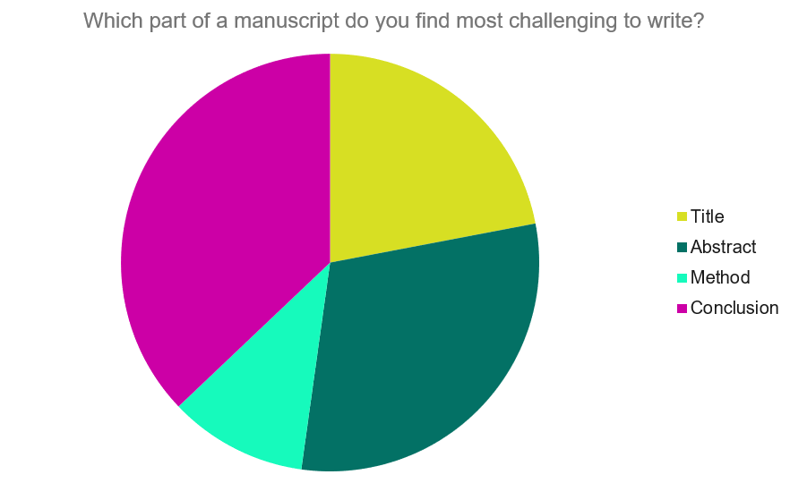
Our Early Career Researcher community tells us that the conclusion is often considered the most difficult aspect of a manuscript to write. To help, this guide provides questions to ask yourself, a basic structure to model your discussion off of and examples from published manuscripts.
Questions to ask yourself:
- Was my hypothesis correct?
- If my hypothesis is partially correct or entirely different, what can be learned from the results?
- How do the conclusions reshape or add onto the existing knowledge in the field? What does previous research say about the topic?
- Why are the results important or relevant to your audience? Do they add further evidence to a scientific consensus or disprove prior studies?
- How can future research build on these observations? What are the key experiments that must be done?
- What is the “take-home” message you want your reader to leave with?
How to structure a discussion
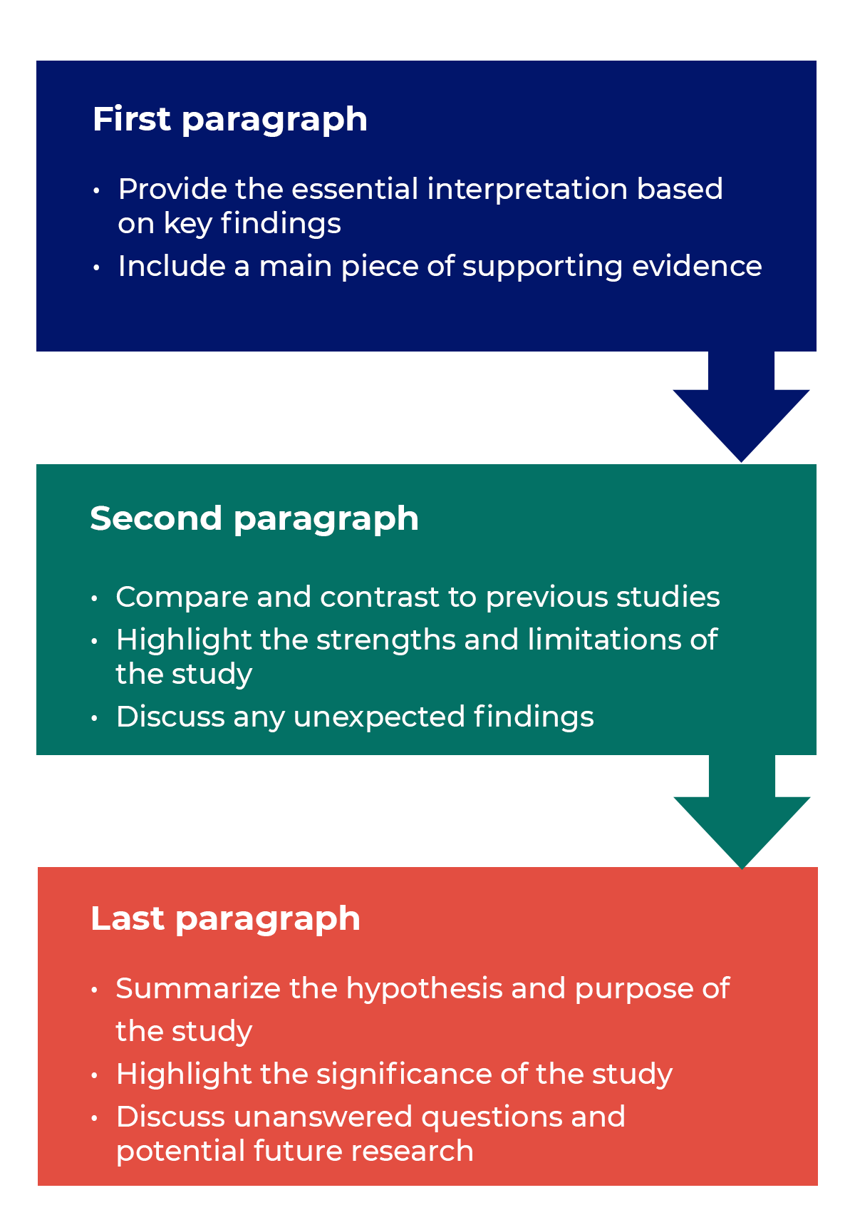
Trying to fit a complete discussion into a single paragraph can add unnecessary stress to the writing process. If possible, you’ll want to give yourself two or three paragraphs to give the reader a comprehensive understanding of your study as a whole. Here’s one way to structure an effective discussion:
Writing Tips
While the above sections can help you brainstorm and structure your discussion, there are many common mistakes that writers revert to when having difficulties with their paper. Writing a discussion can be a delicate balance between summarizing your results, providing proper context for your research and avoiding introducing new information. Remember that your paper should be both confident and honest about the results!

- Read the journal’s guidelines on the discussion and conclusion sections. If possible, learn about the guidelines before writing the discussion to ensure you’re writing to meet their expectations.
- Begin with a clear statement of the principal findings. This will reinforce the main take-away for the reader and set up the rest of the discussion.
- Explain why the outcomes of your study are important to the reader. Discuss the implications of your findings realistically based on previous literature, highlighting both the strengths and limitations of the research.
- State whether the results prove or disprove your hypothesis. If your hypothesis was disproved, what might be the reasons?
- Introduce new or expanded ways to think about the research question. Indicate what next steps can be taken to further pursue any unresolved questions.
- If dealing with a contemporary or ongoing problem, such as climate change, discuss possible consequences if the problem is avoided.
- Be concise. Adding unnecessary detail can distract from the main findings.

Don’t
- Rewrite your abstract. Statements with “we investigated” or “we studied” generally do not belong in the discussion.
- Include new arguments or evidence not previously discussed. Necessary information and evidence should be introduced in the main body of the paper.
- Apologize. Even if your research contains significant limitations, don’t undermine your authority by including statements that doubt your methodology or execution.
- Shy away from speaking on limitations or negative results. Including limitations and negative results will give readers a complete understanding of the presented research. Potential limitations include sources of potential bias, threats to internal or external validity, barriers to implementing an intervention and other issues inherent to the study design.
- Overstate the importance of your findings. Making grand statements about how a study will fully resolve large questions can lead readers to doubt the success of the research.
Snippets of Effective Discussions:
Consumer-based actions to reduce plastic pollution in rivers: A multi-criteria decision analysis approach
Identifying reliable indicators of fitness in polar bears
- How to Write a Great Title
- How to Write an Abstract
- How to Write Your Methods
- How to Report Statistics
- How to Edit Your Work
The contents of the Peer Review Center are also available as a live, interactive training session, complete with slides, talking points, and activities. …
The contents of the Writing Center are also available as a live, interactive training session, complete with slides, talking points, and activities. …
There’s a lot to consider when deciding where to submit your work. Learn how to choose a journal that will help your study reach its audience, while reflecting your values as a researcher…

- Manuscript Preparation
6 Steps to Write an Excellent Discussion in Your Manuscript
- 4 minute read
Table of Contents
The discussion section in scientific manuscripts might be the last few paragraphs, but its role goes far beyond wrapping up. It’s the part of an article where scientists talk about what they found and what it means, where raw data turns into meaningful insights. Therefore, discussion is a vital component of the article.
An excellent discussion is well-organized. We bring to you authors a classic 6-step method for writing discussion sections, with examples to illustrate the functions and specific writing logic of each step. Take a look at how you can impress journal reviewers with a concise and focused discussion section!
Discussion frame structure
Conventionally, a discussion section has three parts: an introductory paragraph, a few intermediate paragraphs, and a conclusion¹. Please follow the steps below:

1.Introduction—mention gaps in previous research¹⁻ ²
Here, you orient the reader to your study. In the first paragraph, it is advisable to mention the research gap your paper addresses.
Example: This study investigated the cognitive effects of a meat-only diet on adults. While earlier studies have explored the impact of a carnivorous diet on physical attributes and agility, they have not explicitly addressed its influence on cognitively intense tasks involving memory and reasoning.
2. Summarizing key findings—let your data speak ¹⁻ ²
After you have laid out the context for your study, recapitulate some of its key findings. Also, highlight key data and evidence supporting these findings.
Example: We found that risk-taking behavior among teenagers correlates with their tendency to invest in cryptocurrencies. Risk takers in this study, as measured by the Cambridge Gambling Task, tended to have an inordinately higher proportion of their savings invested as crypto coins.
3. Interpreting results—compare with other papers¹⁻²
Here, you must analyze and interpret any results concerning the research question or hypothesis. How do the key findings of your study help verify or disprove the hypothesis? What practical relevance does your discovery have?
Example: Our study suggests that higher daily caffeine intake is not associated with poor performance in major sporting events. Athletes may benefit from the cardiovascular benefits of daily caffeine intake without adversely impacting performance.
Remember, unlike the results section, the discussion ideally focuses on locating your findings in the larger body of existing research. Hence, compare your results with those of other peer-reviewed papers.
Example: Although Miller et al. (2020) found evidence of such political bias in a multicultural population, our findings suggest that the bias is weak or virtually non-existent among politically active citizens.
4. Addressing limitations—their potential impact on the results¹⁻²
Discuss the potential impact of limitations on the results. Most studies have limitations, and it is crucial to acknowledge them in the intermediary paragraphs of the discussion section. Limitations may include low sample size, suspected interference or noise in data, low effect size, etc.
Example: This study explored a comprehensive list of adverse effects associated with the novel drug ‘X’. However, long-term studies may be needed to confirm its safety, especially regarding major cardiac events.
5. Implications for future research—how to explore further¹⁻²
Locate areas of your research where more investigation is needed. Concluding paragraphs of the discussion can explain what research will likely confirm your results or identify knowledge gaps your study left unaddressed.
Example: Our study demonstrates that roads paved with the plastic-infused compound ‘Y’ are more resilient than asphalt. Future studies may explore economically feasible ways of producing compound Y in bulk.
6. Conclusion—summarize content¹⁻²
A good way to wind up the discussion section is by revisiting the research question mentioned in your introduction. Sign off by expressing the main findings of your study.
Example: Recent observations suggest that the fish ‘Z’ is moving upriver in many parts of the Amazon basin. Our findings provide conclusive evidence that this phenomenon is associated with rising sea levels and climate change, not due to elevated numbers of invasive predators.
A rigorous and concise discussion section is one of the keys to achieving an excellent paper. It serves as a critical platform for researchers to interpret and connect their findings with the broader scientific context. By detailing the results, carefully comparing them with existing research, and explaining the limitations of this study, you can effectively help reviewers and readers understand the entire research article more comprehensively and deeply¹⁻² , thereby helping your manuscript to be successfully published and gain wider dissemination.
In addition to keeping this writing guide, you can also use Elsevier Language Services to improve the quality of your paper more deeply and comprehensively. We have a professional editing team covering multiple disciplines. With our profound disciplinary background and rich polishing experience, we can significantly optimize all paper modules including the discussion, effectively improve the fluency and rigor of your articles, and make your scientific research results consistent, with its value reflected more clearly. We are always committed to ensuring the quality of papers according to the standards of top journals, improving the publishing efficiency of scientific researchers, and helping you on the road to academic success. Check us out here !
Type in wordcount for Standard Total: USD EUR JPY Follow this link if your manuscript is longer than 12,000 words. Upload
References:
- Masic, I. (2018). How to write an efficient discussion? Medical Archives , 72(3), 306. https://doi.org/10.5455/medarh.2018.72.306-307
- Şanlı, Ö., Erdem, S., & Tefik, T. (2014). How to write a discussion section? Urology Research & Practice , 39(1), 20–24. https://doi.org/10.5152/tud.2013.049

- Manuscript Review
Navigating “Chinglish” Errors in Academic English Writing

A Guide to Crafting Shorter, Impactful Sentences in Academic Writing
You may also like.

Make Hook, Line, and Sinker: The Art of Crafting Engaging Introductions

Can Describing Study Limitations Improve the Quality of Your Paper?

How to Write Clear and Crisp Civil Engineering Papers? Here are 5 Key Tips to Consider

The Clear Path to An Impactful Paper: ②

The Essentials of Writing to Communicate Research in Medicine

Changing Lines: Sentence Patterns in Academic Writing

Path to An Impactful Paper: Common Manuscript Writing Patterns and Structure
Input your search keywords and press Enter.
How to Write the Discussion Section of a Research Paper
The discussion section of a research paper analyzes and interprets the findings, provides context, compares them with previous studies, identifies limitations, and suggests future research directions.
Updated on September 15, 2023

Structure your discussion section right, and you’ll be cited more often while doing a greater service to the scientific community. So, what actually goes into the discussion section? And how do you write it?
The discussion section of your research paper is where you let the reader know how your study is positioned in the literature, what to take away from your paper, and how your work helps them. It can also include your conclusions and suggestions for future studies.
First, we’ll define all the parts of your discussion paper, and then look into how to write a strong, effective discussion section for your paper or manuscript.
Discussion section: what is it, what it does
The discussion section comes later in your paper, following the introduction, methods, and results. The discussion sets up your study’s conclusions. Its main goals are to present, interpret, and provide a context for your results.
What is it?
The discussion section provides an analysis and interpretation of the findings, compares them with previous studies, identifies limitations, and suggests future directions for research.
This section combines information from the preceding parts of your paper into a coherent story. By this point, the reader already knows why you did your study (introduction), how you did it (methods), and what happened (results). In the discussion, you’ll help the reader connect the ideas from these sections.
Why is it necessary?
The discussion provides context and interpretations for the results. It also answers the questions posed in the introduction. While the results section describes your findings, the discussion explains what they say. This is also where you can describe the impact or implications of your research.
Adds context for your results
Most research studies aim to answer a question, replicate a finding, or address limitations in the literature. These goals are first described in the introduction. However, in the discussion section, the author can refer back to them to explain how the study's objective was achieved.
Shows what your results actually mean and real-world implications
The discussion can also describe the effect of your findings on research or practice. How are your results significant for readers, other researchers, or policymakers?
What to include in your discussion (in the correct order)
A complete and effective discussion section should at least touch on the points described below.
Summary of key findings
The discussion should begin with a brief factual summary of the results. Concisely overview the main results you obtained.
Begin with key findings with supporting evidence
Your results section described a list of findings, but what message do they send when you look at them all together?
Your findings were detailed in the results section, so there’s no need to repeat them here, but do provide at least a few highlights. This will help refresh the reader’s memory and help them focus on the big picture.
Read the first paragraph of the discussion section in this article (PDF) for an example of how to start this part of your paper. Notice how the authors break down their results and follow each description sentence with an explanation of why each finding is relevant.
State clearly and concisely
Following a clear and direct writing style is especially important in the discussion section. After all, this is where you will make some of the most impactful points in your paper. While the results section often contains technical vocabulary, such as statistical terms, the discussion section lets you describe your findings more clearly.
Interpretation of results
Once you’ve given your reader an overview of your results, you need to interpret those results. In other words, what do your results mean? Discuss the findings’ implications and significance in relation to your research question or hypothesis.
Analyze and interpret your findings
Look into your findings and explore what’s behind them or what may have caused them. If your introduction cited theories or studies that could explain your findings, use these sources as a basis to discuss your results.
For example, look at the second paragraph in the discussion section of this article on waggling honey bees. Here, the authors explore their results based on information from the literature.
Unexpected or contradictory results
Sometimes, your findings are not what you expect. Here’s where you describe this and try to find a reason for it. Could it be because of the method you used? Does it have something to do with the variables analyzed? Comparing your methods with those of other similar studies can help with this task.
Context and comparison with previous work
Refer to related studies to place your research in a larger context and the literature. Compare and contrast your findings with existing literature, highlighting similarities, differences, and/or contradictions.
How your work compares or contrasts with previous work
Studies with similar findings to yours can be cited to show the strength of your findings. Information from these studies can also be used to help explain your results. Differences between your findings and others in the literature can also be discussed here.
How to divide this section into subsections
If you have more than one objective in your study or many key findings, you can dedicate a separate section to each of these. Here’s an example of this approach. You can see that the discussion section is divided into topics and even has a separate heading for each of them.
Limitations
Many journals require you to include the limitations of your study in the discussion. Even if they don’t, there are good reasons to mention these in your paper.
Why limitations don’t have a negative connotation
A study’s limitations are points to be improved upon in future research. While some of these may be flaws in your method, many may be due to factors you couldn’t predict.
Examples include time constraints or small sample sizes. Pointing this out will help future researchers avoid or address these issues. This part of the discussion can also include any attempts you have made to reduce the impact of these limitations, as in this study .
How limitations add to a researcher's credibility
Pointing out the limitations of your study demonstrates transparency. It also shows that you know your methods well and can conduct a critical assessment of them.
Implications and significance
The final paragraph of the discussion section should contain the take-home messages for your study. It can also cite the “strong points” of your study, to contrast with the limitations section.
Restate your hypothesis
Remind the reader what your hypothesis was before you conducted the study.
How was it proven or disproven?
Identify your main findings and describe how they relate to your hypothesis.
How your results contribute to the literature
Were you able to answer your research question? Or address a gap in the literature?
Future implications of your research
Describe the impact that your results may have on the topic of study. Your results may show, for instance, that there are still limitations in the literature for future studies to address. There may be a need for studies that extend your findings in a specific way. You also may need additional research to corroborate your findings.
Sample discussion section
This fictitious example covers all the aspects discussed above. Your actual discussion section will probably be much longer, but you can read this to get an idea of everything your discussion should cover.
Our results showed that the presence of cats in a household is associated with higher levels of perceived happiness by its human occupants. These findings support our hypothesis and demonstrate the association between pet ownership and well-being.
The present findings align with those of Bao and Schreer (2016) and Hardie et al. (2023), who observed greater life satisfaction in pet owners relative to non-owners. Although the present study did not directly evaluate life satisfaction, this factor may explain the association between happiness and cat ownership observed in our sample.
Our findings must be interpreted in light of some limitations, such as the focus on cat ownership only rather than pets as a whole. This may limit the generalizability of our results.
Nevertheless, this study had several strengths. These include its strict exclusion criteria and use of a standardized assessment instrument to investigate the relationships between pets and owners. These attributes bolster the accuracy of our results and reduce the influence of confounding factors, increasing the strength of our conclusions. Future studies may examine the factors that mediate the association between pet ownership and happiness to better comprehend this phenomenon.
This brief discussion begins with a quick summary of the results and hypothesis. The next paragraph cites previous research and compares its findings to those of this study. Information from previous studies is also used to help interpret the findings. After discussing the results of the study, some limitations are pointed out. The paper also explains why these limitations may influence the interpretation of results. Then, final conclusions are drawn based on the study, and directions for future research are suggested.
How to make your discussion flow naturally
If you find writing in scientific English challenging, the discussion and conclusions are often the hardest parts of the paper to write. That’s because you’re not just listing up studies, methods, and outcomes. You’re actually expressing your thoughts and interpretations in words.
- How formal should it be?
- What words should you use, or not use?
- How do you meet strict word limits, or make it longer and more informative?
Always give it your best, but sometimes a helping hand can, well, help. Getting a professional edit can help clarify your work’s importance while improving the English used to explain it. When readers know the value of your work, they’ll cite it. We’ll assign your study to an expert editor knowledgeable in your area of research. Their work will clarify your discussion, helping it to tell your story. Find out more about AJE Editing.

Adam Goulston, PsyD, MS, MBA, MISD, ELS
Science Marketing Consultant
See our "Privacy Policy"
Ensure your structure and ideas are consistent and clearly communicated
Pair your Premium Editing with our add-on service Presubmission Review for an overall assessment of your manuscript.
Organizing Academic Research Papers: 8. The Discussion
- Purpose of Guide
- Design Flaws to Avoid
- Glossary of Research Terms
- Narrowing a Topic Idea
- Broadening a Topic Idea
- Extending the Timeliness of a Topic Idea
- Academic Writing Style
- Choosing a Title
- Making an Outline
- Paragraph Development
- Executive Summary
- Background Information
- The Research Problem/Question
- Theoretical Framework
- Citation Tracking
- Content Alert Services
- Evaluating Sources
- Primary Sources
- Secondary Sources
- Tertiary Sources
- What Is Scholarly vs. Popular?
- Qualitative Methods
- Quantitative Methods
- Using Non-Textual Elements
- Limitations of the Study
- Common Grammar Mistakes
- Avoiding Plagiarism
- Footnotes or Endnotes?
- Further Readings
- Annotated Bibliography
- Dealing with Nervousness
- Using Visual Aids
- Grading Someone Else's Paper
- How to Manage Group Projects
- Multiple Book Review Essay
- Reviewing Collected Essays
- About Informed Consent
- Writing Field Notes
- Writing a Policy Memo
- Writing a Research Proposal
- Acknowledgements
The purpose of the discussion is to interpret and describe the significance of your findings in light of what was already known about the research problem being investigated, and to explain any new understanding or fresh insights about the problem after you've taken the findings into consideration. The discussion will always connect to the introduction by way of the research questions or hypotheses you posed and the literature you reviewed, but it does not simply repeat or rearrange the introduction; the discussion should always explain how your study has moved the reader's understanding of the research problem forward from where you left them at the end of the introduction.
Importance of a Good Discussion
This section is often considered the most important part of a research paper because it most effectively demonstrates your ability as a researcher to think critically about an issue, to develop creative solutions to problems based on the findings, and to formulate a deeper, more profound understanding of the research problem you are studying.
The discussion section is where you explore the underlying meaning of your research , its possible implications in other areas of study, and the possible improvements that can be made in order to further develop the concerns of your research.
This is the section where you need to present the importance of your study and how it may be able to contribute to and/or fill existing gaps in the field. If appropriate, the discussion section is also where you state how the findings from your study revealed new gaps in the literature that had not been previously exposed or adequately described.
This part of the paper is not strictly governed by objective reporting of information but, rather, it is where you can engage in creative thinking about issues through evidence-based interpretation of findings. This is where you infuse your results with meaning.
Kretchmer, Paul. Fourteen Steps to Writing to Writing an Effective Discussion Section . San Francisco Edit, 2003-2008.
Structure and Writing Style
I. General Rules
These are the general rules you should adopt when composing your discussion of the results :
- Do not be verbose or repetitive.
- Be concise and make your points clearly.
- Avoid using jargon.
- Follow a logical stream of thought.
- Use the present verb tense, especially for established facts; however, refer to specific works and references in the past tense.
- If needed, use subheadings to help organize your presentation or to group your interpretations into themes.
II. The Content
The content of the discussion section of your paper most often includes :
- Explanation of results : comment on whether or not the results were expected and present explanations for the results; go into greater depth when explaining findings that were unexpected or especially profound. If appropriate, note any unusual or unanticipated patterns or trends that emerged from your results and explain their meaning.
- References to previous research : compare your results with the findings from other studies, or use the studies to support a claim. This can include re-visiting key sources already cited in your literature review section, or, save them to cite later in the discussion section if they are more important to compare with your results than being part of the general research you cited to provide context and background information.
- Deduction : a claim for how the results can be applied more generally. For example, describing lessons learned, proposing recommendations that can help improve a situation, or recommending best practices.
- Hypothesis : a more general claim or possible conclusion arising from the results [which may be proved or disproved in subsequent research].
III. Organization and Structure
Keep the following sequential points in mind as you organize and write the discussion section of your paper:
- Think of your discussion as an inverted pyramid. Organize the discussion from the general to the specific, linking your findings to the literature, then to theory, then to practice [if appropriate].
- Use the same key terms, mode of narration, and verb tense [present] that you used when when describing the research problem in the introduction.
- Begin by briefly re-stating the research problem you were investigating and answer all of the research questions underpinning the problem that you posed in the introduction.
- Describe the patterns, principles, and relationships shown by each major findings and place them in proper perspective. The sequencing of providing this information is important; first state the answer, then the relevant results, then cite the work of others. If appropriate, refer the reader to a figure or table to help enhance the interpretation of the data. The order of interpreting each major finding should be in the same order as they were described in your results section.
- A good discussion section includes analysis of any unexpected findings. This paragraph should begin with a description of the unexpected finding, followed by a brief interpretation as to why you believe it appeared and, if necessary, its possible significance in relation to the overall study. If more than one unexpected finding emerged during the study, describe each them in the order they appeared as you gathered the data.
- Before concluding the discussion, identify potential limitations and weaknesses. Comment on their relative importance in relation to your overall interpretation of the results and, if necessary, note how they may affect the validity of the findings. Avoid using an apologetic tone; however, be honest and self-critical.
- The discussion section should end with a concise summary of the principal implications of the findings regardless of statistical significance. Give a brief explanation about why you believe the findings and conclusions of your study are important and how they support broader knowledge or understanding of the research problem. This can be followed by any recommendations for further research. However, do not offer recommendations which could have been easily addressed within the study. This demonstrates to the reader you have inadequately examined and interpreted the data.
IV. Overall Objectives
The objectives of your discussion section should include the following: I. Reiterate the Research Problem/State the Major Findings
Briefly reiterate for your readers the research problem or problems you are investigating and the methods you used to investigate them, then move quickly to describe the major findings of the study. You should write a direct, declarative, and succinct proclamation of the study results.
II. Explain the Meaning of the Findings and Why They are Important
No one has thought as long and hard about your study as you have. Systematically explain the meaning of the findings and why you believe they are important. After reading the discussion section, you want the reader to think about the results [“why hadn’t I thought of that?”]. You don’t want to force the reader to go through the paper multiple times to figure out what it all means. Begin this part of the section by repeating what you consider to be your most important finding first.
III. Relate the Findings to Similar Studies
No study is so novel or possesses such a restricted focus that it has absolutely no relation to other previously published research. The discussion section should relate your study findings to those of other studies, particularly if questions raised by previous studies served as the motivation for your study, the findings of other studies support your findings [which strengthens the importance of your study results], and/or they point out how your study differs from other similar studies. IV. Consider Alternative Explanations of the Findings
It is important to remember that the purpose of research is to discover and not to prove . When writing the discussion section, you should carefully consider all possible explanations for the study results, rather than just those that fit your prior assumptions or biases.
V. Acknowledge the Study’s Limitations
It is far better for you to identify and acknowledge your study’s limitations than to have them pointed out by your professor! Describe the generalizability of your results to other situations, if applicable to the method chosen, then describe in detail problems you encountered in the method(s) you used to gather information. Note any unanswered questions or issues your study did not address, and.... VI. Make Suggestions for Further Research
Although your study may offer important insights about the research problem, other questions related to the problem likely remain unanswered. Moreover, some unanswered questions may have become more focused because of your study. You should make suggestions for further research in the discussion section.
NOTE: Besides the literature review section, the preponderance of references to sources in your research paper are usually found in the discussion section . A few historical references may be helpful for perspective but most of the references should be relatively recent and included to aid in the interpretation of your results and/or linked to similar studies. If a study that you cited disagrees with your findings, don't ignore it--clearly explain why the study's findings differ from yours.
V. Problems to Avoid
- Do not waste entire sentences restating your results . Should you need to remind the reader of the finding to be discussed, use "bridge sentences" that relate the result to the interpretation. An example would be: “The lack of available housing to single women with children in rural areas of Texas suggests that...[then move to the interpretation of this finding].”
- Recommendations for further research can be included in either the discussion or conclusion of your paper but do not repeat your recommendations in the both sections.
- Do not introduce new results in the discussion. Be wary of mistaking the reiteration of a specific finding for an interpretation.
- Use of the first person is acceptable, but too much use of the first person may actually distract the reader from the main points.
Analyzing vs. Summarizing. Department of English Writing Guide. George Mason University; Discussion . The Structure, Format, Content, and Style of a Journal-Style Scientific Paper. Department of Biology. Bates College; Hess, Dean R. How to Write an Effective Discussion. Respiratory Care 49 (October 2004); Kretchmer, Paul. Fourteen Steps to Writing to Writing an Effective Discussion Section . San Francisco Edit, 2003-2008; The Lab Report . University College Writing Centre. University of Toronto; Summary: Using it Wisely . The Writing Center. University of North Carolina; Schafer, Mickey S. Writing the Discussion . Writing in Psychology course syllabus. University of Florida; Yellin, Linda L. A Sociology Writer's Guide. Boston, MA: Allyn and Bacon, 2009.
Writing Tip
Don’t Overinterpret the Results!
Interpretation is a subjective exercise. Therefore, be careful that you do not read more into the findings than can be supported by the evidence you've gathered. Remember that the data are the data: nothing more, nothing less.
Another Writing Tip
Don't Write Two Results Sections!
One of the most common mistakes that you can make when discussing the results of your study is to present a superficial interpretation of the findings that more or less re-states the results section of your paper. Obviously, you must refer to your results when discussing them, but focus on the interpretion of those results, not just the data itself.
Azar, Beth. Discussing Your Findings. American Psychological Association gradPSYCH Magazine (January 2006)
Yet Another Writing Tip
Avoid Unwarranted Speculation!
The discussion section should remain focused on the findings of your study. For example, if you studied the impact of foreign aid on increasing levels of education among the poor in Bangladesh, it's generally not appropriate to speculate about how your findings might apply to populations in other countries without drawing from existing studies to support your claim. If you feel compelled to speculate, be certain that you clearly identify your comments as speculation or as a suggestion for where further research is needed. Sometimes your professor will encourage you to expand the discussion in this way, while others don’t care what your opinion is beyond your efforts to interpret the data.
- << Previous: Using Non-Textual Elements
- Next: Limitations of the Study >>
- Last Updated: Jul 18, 2023 11:58 AM
- URL: https://library.sacredheart.edu/c.php?g=29803
- QuickSearch
- Library Catalog
- Databases A-Z
- Publication Finder
- Course Reserves
- Citation Linker
- Digital Commons
- Our Website
Research Support
- Ask a Librarian
- Appointments
- Interlibrary Loan (ILL)
- Research Guides
- Databases by Subject
- Citation Help
Using the Library
- Reserve a Group Study Room
- Renew Books
- Honors Study Rooms
- Off-Campus Access
- Library Policies
- Library Technology
User Information
- Grad Students
- Online Students
- COVID-19 Updates
- Staff Directory
- News & Announcements
- Library Newsletter
My Accounts
- Interlibrary Loan
- Staff Site Login
FIND US ON
Page Contents
- 1.1 Research Answer
- 1.2 Key Findings
- 1.3 Interpretations
- 1.4 Comparison to Other Studies
- 1.5 Acknowledgement of Limitations
- 1.6 Recommendations for Future Research
- 2 Six Key Components of the Discussion Section – An Example
- 3 Questions to Help You Interpret Your Results
- 4 Reflective Exercise – Interpreting Your Results
- 5 Common Structure of the Discussion Section
- 6.1 Tip 1 – Include Specific Limitations
- 6.2 Tip 2 – Conclude with Your Contributions
- 6.3 Tip 3 – Use Signal Phrases
- 6.4 Tip 4 – Choose Verbs Carefully
- 8 Conclusion
Writing the Discussion
The Discussion section is widely recognized as the most challenging part of the research article to write. But it’s also the most rewarding section in many ways because it’s where you get to say what your findings mean and why they matter. It’s where you get to talk about your own contributions to the research. Before you start writing your discussion, think critically about your data so that you can share your research story with your reader.
The Discussion section of a research article answers,

The six main components of the discussion section that will help you answer these questions for your reader are

The following list provides a brief overview of each of these components. The Common Structure of the Discussion section provides more details about how these components are integrated and developed within the paragraphs of the Discussion.
Research Answer

Start your Discussion by explicitly answering your research question. If you had a hypothesis, indicate whether or not it was supported. In some cases, you may wish to remind your reader of the research question before providing your research answer.
Key Findings
Provide an overview of your major findings before offering your specific interpretations and comparisons to other studies.

Interpretations

Explain what your results mean and make any claims based on your results. Ensure that you ground all your claims in evidence
Comparison to Other Studies
Compare your findings to those of other studies. Within the Discussion, interpretations and comparisons to other studies are often integrated. As you interpret your findings, you’ll indicate how they compare to existing research, and what the similarities or differences suggest. You’ll repeat this pattern as you move through your findings.

Acknowledgement of Limitations

Highlight the specific limitations of your study to demonstrate your awareness of potential gaps, acknowledge methodological drawbacks, and anticipate potential questions or criticism.
Recommendations for Future Research
Indicate what future researchers can do to build upon your research findings and take the research further.

Six Key Components of the Discussion Section – An Example
This video illustrates the six key components of the discussion section in a scientific research article by examining excerpts from a fictional research article about varroa mites in honeybee colonies.
NOTE: For educational purposes, we’ve created fictional excerpts that resemble passages from scientific research articles. The fictional examples are intended to illustrate writing techniques and are not designed to teach scientific content. Please note that the scientific content and data in this video is fictional.
[Background sounds of bees buzzing and birds chirping]
To illustrate the six key components of the discussion section, we’ll examine excerpts from a fictional scientific research article about varroa mites in honeybee colonies.
The writer starts their discussion with an ANSWER to their RESEARCH QUESTION by noting which two natural chemical treatments are effective. [A laptop displays the following voiceover text on screen.] They state, “Our findings indicate that formic acid strips and oxalic acid trickling are effective natural chemical treatments for reducing the presence of varroa mites in honeybee colonies.”
Next, the writer provides a summary of their KEY FINDINGS by focusing on which of the two treatments are most effective: [A laptop displays the following voiceover text on screen.] “Formic acid treatment was the most effective treatment for reducing the presence of varroa mites in honeybee colonies. This finding suggests that this treatment is an effective option for Ontario beekeepers.”
After providing the key findings, the writer begins to INTERPRET their INDIVIDUAL FINDINGS. The writer provides explanations for the similarities and differences that they observe. [A laptop displays the following voiceover text on screen.] For example, they note that, “Although oxalic acid is stronger than formic acid, formic acid strips were 14% more effective than oxalic acid trickling in reducing varroa mite populations. One reason for this observed difference may be that formic acid can penetrate the wax of the brood chamber whereas oxalic acid cannot.”
The writer also COMPARES their findings to OTHER STUDIES. They state, “Our findings with respect to natural chemical treatments are similar to those of Buzz et al. (2021). Buzz et al. (2021) compared the efficacy of sucrocide spray treatment to formic acid treatment and found that formic acid treatment was more effective for reducing mite populations. These similarities suggest that formic acid treatments are an effective option that beekeepers can use to protect their colonies.”
Note here that the writer moves beyond stating that their research is similar to that of others. [Laptop screen showing the text “These similarities suggest that formic acid treatments are an effective option that beekeepers can use to protect their colonies.”] They also indicate what these similarities suggest about the results.
In addition to highlighting what they found, the writer also ACKNOWLEDGES their LIMITATIONS. [A laptop displays the following voiceover text on screen.] They note, “As our study took place during a single season, we did not have the opportunity to determine how temperature impacts treatment efficacy.”
[Text on screen “SUGGEST FUTURE RESEARCH.] The writer concludes by indicating how this research can be addressed by future researchers, noting, “Further research is needed to determine how seasonal temperature impacts the efficacy of treatment types.”
Questions to Help You Interpret Your Results
Every claim that you make in your Discussion section must be grounded in evidence. Ensure that you understand your results thoroughly and present them effectively. These are some questions that you can ask yourself to determine what your findings mean and what you plan to write about them:

Examine the results from your study and consider how they relate to your research question or your hypotheses if you’re doing hypothesis-driven research. Which results did you expect? Which results did you not expect?

Think about how your results compare to the literature that you explored for your introduction, research proposal, or literature reviews. How do your results compare to existing studies? What are the similarities? What are the differences?

Although indicating how your research is similar to or different from existing research is important, you need to move beyond these statements to provide your own interpretations as well. Consider what these similarities and differences suggest. What do similarities between your results and those of other researchers mean? What might be some reasons for any differences that you’ve observed? Are there any interesting implications to note?
These questions will help you approach your data with a critical eye and map out the possible interpretations. Consider the following advice from Joshua Schimel’s Writing Science: How to Write Papers That Get Cited and Proposals That Get Funded (2012).
“What might that shoulder on the spectrum mean? If that nonsignificant treatment effect were real, what would that say about your system? Is that outlier a flag for something you hadn’t thought about but may be important? Overinterpret your data wildly, and consider what they might mean at those farthest fringes. Explore the possibilities and develop the story expansively. Then, take Occam’s razor and slash away to find the simple core” (Schimel 2012, p. 12).
As Schimel emphasizes in this passage, interpreting your data critically is key to telling the story of your research findings. A starting place is to consider all the possibilities for what your research could mean to ensure that you don’t miss possible interpretations. Once you’ve completed this exercise, consider the principle of Occam’s razor – the idea that the simplest explanation is usually the best one – to ensure that you’re not over interpreting your data. Once you start writing, focus your Discussion on the interpretations that you can provide specific evidence to support.
Reflective Exercise – Interpreting Your Results
The following “Interpreting Your Results – Worksheet” is a tool designed to guide your critical reflection and writing process. Use the table on this worksheet to help you interpret your results and discuss your research findings.
NOTE: You can view this worksheet online, but you can also download it below as an accessible screen reader document.
Download PDF (Interpreting Your Results – Worksheet)
This table is divided into four columns:
- Column One: Describe a result from your study.
- Column Two: Explain what your result indicates in a direct way. In other words, what would experts who look at this result logically conclude from it?
- Column Three: Consider what claims you could make about the result. In other words, what are your specific thoughts and interpretations about what the data could mean?
- Column Four: Note any questions that you still have about your result. These could be questions that you could answer by revisiting the literature in your field, or they could be questions that future researchers should consider.
Common Structure of the Discussion Section
In the Discussion section, writers typically move from a specific statement of research findings to the broad implications of the work. This movement is the opposite trajectory of what you typically see in an Introduction section, where the image of a funnel often represents how the writer will move from the broad area of research to the narrow, specific research question. The opposite image – that of a pyramid – is useful for the Discussion section. However, a Discussion section is not simply a backwards Introduction. In the Discussion, writers start with the answer to their specific research question and then move outward to discuss the broad implications of their work.

The following structure is a common one that you will find in the Discussion section of many research articles in the sciences.
Opening Paragraph: Provide your research answer and state your key findings.
Body Paragraphs: Offer your interpretations and comparisons to other studies. Carefully consider the order in which you present your body paragraphs. Often, writers will start with the findings that are most central to the research question and then move into findings that are less critical.
There are two main ways to organize body paragraphs in the Discussion section.

Image description: Two images showing different structures for body paragraphs. The first image [Option 1] has a large box with the text “My result indicates…” and a smaller box beneath with text “… and this … is how my result compares to other literature.” The second image [Option 2] has a large box with the text “The existing literature shows…” and a smaller box beneath with text “… and this … is how my result fits into the literature.”
Option 1 is to discuss the meaning of a result and then compare it to the existing literature. Option 2 is to write about the relevant literature and then discuss how your results fit in. Both options are valid, and you’ll see both in published research articles.
However, option 1, where you start the paragraph with your own findings, more effectively highlights your research contributions. The Discussion section is the part of your article where you get to highlight what your results mean and why your findings are important. Option 2 makes other researchers’ work the primary focus of your Discussion, and then you risk burying your own contributions, and your Discussion section could read like a literature review. It is critical to foreground your own research contributions so that readers know why your research is important.
Concluding Paragraph: Provide your acknowledgment of limitations and recommendations for future research . Note the broader implications of your study by returning to the major topic that you introduced in your opening paragraph of the research article.
Note: Sometimes the concluding paragraph of the Discussion will appear under a separate “Conclusion” heading. A conclusion notes the contribution of the study to the field and indicates what researchers should explore next. When you’re reading articles in your target journal, take note of whether these journals include a separate Conclusion section.
Writing Tips
Below are four writing tips for writing your Discussion section:
Tip 1 – Include Specific Limitations
When you’re writing your Discussion section, you may feel hesitant to include limitations. You may worry that by mentioning a limitation, you’ve brought it to the reader’s attention when the reader wouldn’t have thought of it otherwise. You may worry that by drawing attention to a limitation, you’re making your research look weak.
While some readers may not notice your limitations until you point them out, overall, academics are trained to read critically. Academics are often thinking about potential limitations as part of their critical reading practice.
While you want to indicate how your research contributes to the field, you want to be cautious about overestimating or overstating your research findings. Because readers are trained to read critically, you’ll want to be thinking about potential objections or potential questions about your work.
Write these potential objections or questions down, consider which ones are most relevant, and acknowledge them as part of writing your limitations. Acknowledging limitations qualifies your contribution in a meaningful way and strengthens your writing.
Tip 2 – Conclude with Your Contributions
In the final paragraph of your Discussion section, reiterate your major contributions to the field of research that you introduced in your introduction section. Ending with a statement of your contributions is stronger than ending with a description of your limitations. Although stating limitations is important, you also want to ensure that your reader knows why your research matters and how it contributes to the field. Information that you place at the end of a section creates emphasis. This is the information that the reader is left with, so end on a strong note.
Tip 3 – Use Signal Phrases
Throughout your Discussion, use signal phrases to highlight your key components.
Here are a couple of examples of signal phrases that you can use. “Our findings indicate that” is a simple phrase that you can use to provide your answer to the research question. “One limitation of this study is” is a direct way that you can acknowledge your limitations. These phrases are easy for your reader to spot and also easy for reviewers and editors to see.
When you’re reading other research articles, take a look at where and how writers are signalling their key components so that you can get a sense what techniques work well for you as a reader.
Tip 4 – Choose Verbs Carefully
In the Discussion section, choose verbs that accurately reflect your level of certainty about your findings. In research writing, you’ll often see verbs like “suggests,” “indicates,” and “shows.” You’ll rarely see the verb “proves” because of the extreme level of certainty associated with this word.
Within the sciences, you’re working within a tradition where you’re incrementally building upon the findings of others, and within this tradition, it can be perceived as arrogant at best or actively dangerous at worst to overstate your research findings, particularly if you’re making claims that your data don’t support.
Modal verbs like “can” and “may” qualify our level of certainty. Outside of academia, modal verbs can sometimes be viewed negatively because they can be seen to undermine the force of our statements or make our claims seem uncertain or weak. However, within research writing, modal verbs are more acceptable because we’re being cautious.
It’s important to be cautious because people may make major policy decisions or undertake particular treatment plans because of your research, so choose verbs that accurately reflect your certainty and consider that using modal verbs to qualify your level of certainty can help you advance ideas carefully while acknowledging a continued need for research. However, keep in mind that modal verbs can also be overused, so think carefully before you use one. If your findings are novel, don’t let a modal verb detract from that.
In the Discussion section, choose your verbs carefully and revise if necessary. Word choice matters.
In some disciplines within the sciences, Results and Discussion sections are combined into a single section. For example, combined Results and Discussion sections are common in research articles in engineering journals. Combined sections are also common in shorter pieces of writing and more visual mediums, such as scientific posters.
Scientists who carry out modeling and simulations will often combine their Results and Discussion sections. This approach allows them to tell a more cohesive story as they can discuss the significance of each model or simulation right after presenting the results.
If you’re opting to combine your Results and Discussion section, consider using subheadings throughout the section to make it more navigable for your reader. A combined section will often still move from presenting results at the beginning to interpreting results at the end.
When you’re investigating your target journal, explore whether writers typically combine their Results and Discussion section. If you see instances of both separate and combined sections, consider which approach will allow you to tell a cohesive story about your research while also clearly differentiating between the results of the research and your interpretations of those results.
There are many ways to approach writing the Discussion section for a mixed-methods study. Analyzing examples of mixed methods research articles from your field is a good starting place for approaching this writing task.
If you’ve conducted a mixed-methods study and have both quantitative and qualitative data, the Discussion can be a good place to provide an integrated interpretation of any relationships between both data sets. For example, you could use the following approach in a paragraph:
1) Provide a topic sentence that introduces the subject of the paragraph.
2) Discuss a quantitative result and consider comparing it to any relevant literature.
3) Discuss a qualitative result and consider comparing it to any relevant literature.
4) Explain what the quantitative and qualitative results mean when we consider them together.
Our findings show that new beekeepers in Ontario have limited familiarity with using formic acid strips to control varroa mites. In response to our survey question of whether they had applied formic acid strips to their colonies, 70% of new beekeepers noted they had used this method within the past two years, indicating that the treatment method itself is widely known. However, in our interviews, new beekeepers noted that although they had applied formic acid strips, they had received little education and training in how to do so successfully, and the method consequently was not effective in controlling the varroa mite population of their colonies. These findings suggest that new beekeepers lack a developed understanding of how to apply formic acid strips effectively despite their widespread awareness of this treatment method and willingness to use it on their colonies.
For additional examples of Discussion paragraphs that integrate quantitative and qualitative findings, see Bronstein and Kovacs (2013) .
Now that you’ve identified the key components of the Discussion section, you can fill out the Discussion page of your research article map , Next, you’re ready to write your abstract and title.

The content of this page was created by Dr. Jodie Salter and Dr. Sarah Gibbons.
About this website


Chapter 6: Discussion/Conclusion Section(s)
Discussion/Conclusion Goal 2: Framing Principal Findings
The next goal in the Discussion and Conclusion section is Framing Principal Findings. The function of this goal is to discuss and establish the meaning of your research results. The name Framing Principal Findings comes from the aim of positioning the outcomes of your study in a way that the reader can understand — typically, within the already existing literature on the topic.
There are four strategies that can be used to achieve the communicative aims of Goal 2. Note that these are primarily associated with discussion of results rather than drawing conclusions about the entirety of the research.
Strategies for Discussion/Conclusion Communicative Goal 2: Framing Principal Findings
- Accounting for results
- Explicating results
- Relating to expectations
- Addressing limitations
Discussion/Conclusion Goal 2 Strategy: Accounting for Results
Accounting for results is a way for you to reflect on or further consider the findings of your study. This could be accomplished with or without referencing previous works. You can use this strategy to clarify what may have contributed to, caused, or otherwise affected the results or outcomes. You may also want to suggest reasons or hypotheses that could account for certain findings, and justify the nature of results.
Here are two examples of how you can accomplish this strategy, with strategy-specific language in bold :
- These changes are most probably related to modifications in N uptake into aap2 mesophyll cells, as suggested by upregulation of amino acid importer LHT1 (Figure 7A) and increased uptake of 14C-label. [1]
- That multiple genes in this pathway are underexpressed in hybrids of both species pairs perhaps is a cause or consequence of their sterility. [2]
The Academic Phrasebank website offers a wealth of sentence starters useful to writers in this stage of reporting their research:
General comments and explanations:
- A possible explanation for this might be that …
- Another possible explanation for this is that …
- This result may be explained by the fact that …
- There are, however, other possible explanations.
- These relationships may partly be explained by …
- There are several possible explanations for this result.
- These results are likely to be related to …
- It seems possible that these results are due to …
Reporting inconsistent, counterintuitive, or contradictory findings:
- These differences can be explained in part by …
- This inconsistency may be due to …
- This discrepancy could be attributed to …
- This rather contradictory result may be due to …
- The observed increase in X could be attributed to …
- It is difficult to explain this result, but it might be related to …
- The possible interference of X cannot be ruled out.
- Differences between X and Y may have influenced …
- These possible sources of error could have affected …
- There are two likely causes for the differences between …
- The reason for this is not clear but it may have something to do with …
- Since this difference has not been found elsewhere it is probably not due to …
Discussion/Conclusion Goal 2 Strategy: Explicating Results
The expli cating results strategy explains the reported results in the context of the study and/or in a broader context of the discipline. Here is where you consider your results and their implications. Your readers, of course, will develop their own interpretations or explanations for why the findings turned out as they did; however, you will want to guide their views as you make an argument explaining your own reasoning.
Here are a couple of examples from published articles in high-impact journals:
- We must therefore conclude , to use the terminology of Goodlad et al. (1979), that the content standards, as formulated by the relevant official bodies, have not been properly incorporated into the formal curriculum, despite their authoritative national status. [3]
- This research also showed that the effectiveness of rotations at reducing the weed seedbank was dependent on the specific crop that initiated the rotations (Fig. 1-3). [4]
The Academic Phrasebank website provides this list of suggested ways to report findings that are related to previous research:
Reporting findings in line with previous research:
- Several reports have shown that …
- As mentioned in the literature review, …
- Prior studies that have noted the importance of …
- Very little was found in the literature on the question of …
- Previous studies evaluating X observed inconsistent results on whether …
- A strong relationship between X and Y has been reported in the literature.
- In reviewing the literature, no data was found on the association between X and Y.
- These factors may explain the relatively good correlation between X and Y.
- It may be that these participants benefitted from …
- The observed correlation between X and Y might be explained in this way: …
Discussion/Conclusion Goal 2 Strategy: Relating to Expectations
Relating to expectations reasons about the researchers’ anticipated or unanticipated findings and/or observations. You can use this strategy to point out expected or unexpected results, express your attitudes about the results, often concerning surprise or unsatisfactory findings, and connect findings to initial hypotheses (i.e., to describe how findings were or were not confirmed, as seen in the following examples):
- Hypothesis 1 is verified by unchanged oil content values in rain-fed plot. [5]
- Interestingly, relative to inulin the dimer exhibited higher activity than the tetramer [35]. [6]
The Academic Phrasebank website recommends these phrases that are in line with using this strategy:
- Surprisingly, X was found to …
- What is surprising is that …
- One unanticipated finding was that …
- Surprisingly, no differences were found in …
- This finding was unexpected and suggests that …
- It is somewhat surprising that no X was noted in this condition …
- Contrary to expectations, this study did not find a significant difference between …
- However, the observed difference between X and Y in this study was not significant.
- However, the ANOVA (one way) showed that these results were not statistically significant.
Discussion/Conclusion Goal 2 Strategy: Addressing Limitations
Addressing limitations is used to evaluate the study by pointing out shortcomings and/or minimizing deficiencies in the research.
The following are examples of how you can realize this strategy, with specific language for expressing these limitations in bold .
- However, the study area spans an overly large region encompassing several climatic zones, which calls into question whether chronologies can simply be averaged. [7]
- Our study was not free of problems, however . For instance, the overall rate of participation in sending feedback by e-mail was just over 50% of all participants (23 corrective feedback e-mails to 44 transcripts in English sessions and 23 corrective feedback e-mails to 43 transcripts in Japanese sessions). [8]
It’s important to note that research reports do not provide information without taking a stance toward that information. Researchers may indicate more or less certainty about their interpretations, and this is especially true of Addressing Limitations. While it’s important, of course, to point to the shortcomings of your study (as no study is perfect), it’s important not to over-emphasize the deficiencies in your research. So, you may want to highlight a contribution that your study makes while simultaneously acknowledging some aspect that was less than ideal. In that case, you would want to hedge or boost accordingly, as showing in these sentence starters from the Academic Phrasebank website :
Sometimes it isn’t necessary to hedge or boost, but rather to simply present the information as neutrally as possible.
- It could be argued that the positive results were due to …
- Although the current study is based on a small sample of participants, the findings suggest …
- While this study did not confirm X, it did …
- Notwithstanding these limitations, the study suggests that …
- Despite its exploratory nature, this study offers some insight into …
- A note of caution is due here since …
- Another source of uncertainty is …
- These results, therefore, need to be interpreted with caution.
- In observational studies, there is a potential for bias from …
- It is important to bear in mind the possible bias in these responses.
Key Takeaways
Goal 2 of writing the Discussion/Conclusion section is related to Frame Principal Findings . There are four possible strategies that you can use to accomplish this goal:
- Accounting for results, and/or
- Explicating results, and/or
- Relating to expectations, and/or
- Addressing limitations.
Remember: It isn’t necessary to include all of these strategies — they are simply possibilities for reaching the goal.
- Zhang, L., Tan, Q., Lee, R., Trethewy, A., Lee, Y. H., & Tegeder, M. (2010). Altered xylem-phloem transfer of amino acids affects metabolism and leads to increased seed yield and oil content in Arabidopsis. The Plant Cell , 22 (11), 3603-3620. ↵
- Noor, M. A. (2005). Patterns of evolution of genes disrupted in expression in Drosophila species hybrids. Genetics Research , 85 (2), 119-125. ↵
- Fukkink, R. G. (2010). Missing pages? A study of textbooks for Dutch early childhood teacher education. Teaching and Teacher Education , 26 (3), 371-376. ↵
- Teasdale, J. R., Mangum, R. W., Radhakrishnan, J., & Cavigelli, M. A. (2004). Weed seedbank dynamics in three organic farming crop rotations. Agronomy Journal , 96 (5), 1429-1435. ↵
- Bedbabis, S., Rouina, B. B., & Boukhris, M. (2010). The effect of waste water irrigation on the extra virgin olive oil quality from the Tunisian cultivar Chemlali. Scientia Horticulturae , 125 (4), 556-561.. ↵
- Artyukhov, V. G., Kovaleva, T. A., Kholyavka, M. G., Bityutskaya, L. A., Grechkina, M. V., & Obraztsova, T. B. (2009). Study of the oligomeric structure and some physicochemical properties of inulinase from Kluyveromyces marxianus Y-303. Biophysics, 54 (6), 675-680. ↵
- Black, B. A., Copenheaver, C. A., Frank, D. C., Stuckey, M. J., & Kormanyos, R. E. (2009). Multi-proxy reconstructions of northeastern Pacific sea surface temperature data from trees and Pacific geoduck. Palaeogeography, Palaeoclimatology, Palaeoecology , 278 (1-4), 40-47. ↵
- Bower, J., & Kawaguchi, S. (2011). Negotiation of meaning and corrective feedback in Japanese/English eTandem. Language Learning & Technology , 15 (1), 41-71. ↵
Preparing to Publish Copyright © 2023 by Sarah Huffman; Elena Cotos; and Kimberly Becker is licensed under a Creative Commons Attribution-NonCommercial-ShareAlike 4.0 International License , except where otherwise noted.

Discussion Vs. Conclusion: Know the Difference Before Drafting Manuscripts
The discussion section of your manuscript can be one of the hardest to write as it requires you to think about the meaning of the research you have done. An effective discussion section tells the reader what your study means and why it is important. In this article, we will cover some pointers for writing clear/well-organized discussion and conclusion sections and discuss what should NOT be a part of these sections.
What Should be in the Discussion Section?
Your discussion is, in short, the answer to the question “what do my results mean?” The discussion section of the manuscript should come after the methods and results section and before the conclusion. It should relate back directly to the questions posed in your introduction, and contextualize your results within the literature you have covered in your literature review . In order to make your discussion section engaging, you should include the following information:
- The major findings of your study
- The meaning of those findings
- How these findings relate to what others have done
- Limitations of your findings
- An explanation for any surprising, unexpected, or inconclusive results
- Suggestions for further research
Your discussion should NOT include any of the following information:
- New results or data not presented previously in the paper
- Unwarranted speculation
- Tangential issues
- Conclusions not supported by your data
Related: Avoid outright rejection with a well-structured manuscript. Check out these resources and improve your manuscript now!
How to Make the Discussion Section Effective?
There are several ways to make the discussion section of your manuscript effective, interesting, and relevant. Hear from one of our experts on how to structure your discussion section and distinguish it from the results section:
Now that we have listened to how to approach writing a discussion section, let’s delve deeper into some essential tips with a few examples:
- Most writing guides recommend listing the findings of your study in decreasing order of their importance. You would not want your reader to lose sight of the key results that you found. Therefore, put the most important finding front and center. Example: Imagine that you conduct a study aimed at evaluating the effectiveness of stent placement in patients with partially blocked arteries. You find that despite this being a common first-line treatment, stents are not effective for patients with partially blocked arteries. The study also discovers that patients treated with a stent tend to develop asthma at slightly higher rates than those who receive no such treatment.
Which sentence would you choose to begin your discussion? Our findings suggest that patients who had partially blocked arteries and were treated with a stent as the first line of intervention had no better outcomes than patients who were not given any surgical treatments. Our findings noted that patients who received stents demonstrated slightly higher rates of asthma than those who did not. In addition, the placement of a stent did not impact their rates of cardiac events in a statistically significant way.
If you chose the first example, you are correct!
- If you are not sure which results are the most important, go back to your research question and start from there. The most important result is the one that answers your research question.
- It is also necessary to contextualize the meaning of your findings for the reader. What does previous literature say, and do your results agree? Do your results elaborate on previous findings, or differ significantly?
- In our stent example, if previous literature found that stents were an effective line of treatment for patients with partially blocked arteries, you should explore why your interpretation seems different in the discussion section. Did your methodology differ? Was your study broader in scope and larger in scale than the previous studies? Were there any limitations to previous studies that your study overcame? Alternatively, is it possible that your own study could be incorrect because of some difficulties you had in carrying it out? The discussion section should narrate a coherent story to the target audience.
- Finally, remember not to introduce new ideas/data, or speculate wildly on the possible future implications of your study in the discussion section. However, considering alternative explanations for your results is encouraged.

Avoiding Confusion in your Conclusion!
Many writers confuse the information they should include in their discussion with the information they should place in their conclusion. One easy way to avoid this confusion is to think of your conclusion as a summary of everything that you have said thus far. In the conclusion section, you remind the reader of what they have just read. Your conclusion should:
- Restate your hypothesis or research question
- Restate your major findings
- Tell the reader what contribution your study has made to the existing literature
- Highlight any limitations of your study
- State future directions for research/recommendations
Your conclusion should NOT:
- Introduce new arguments
- Introduce new data
- Fail to include your research question
- Fail to state your major results
An appropriate conclusion to our hypothetical stent study might read as follows:
In this study, we examined the effectiveness of stent placement. We compared the patients with partially blocked arteries to those with non-surgical interventions. After examining the five-year medical outcomes of 19,457 patients in the Greater Dallas area, our statistical analysis concluded that the placement of a stent resulted in outcomes that were no better than non-surgical interventions such as diet and exercise. Although previous findings indicated that stent placement improved patient outcomes, our study followed a greater number of patients than those in major studies conducted previously. It is possible that outcomes would vary if measured over a ten or fifteen year period. Future researchers should consider investigating the impact of stent placement in these patients over a longer period (five years or longer). Regardless, our results point to the need for medical practitioners to reconsider the placement of a stent as the first line of treatment as non-surgical interventions may have equally positive outcomes for patients.
Did you find the tips in this article relevant? What is the most challenging portion of a research paper for you to write? Let us know in the comments section below!

This is the most stunning and self-instructional site I have come across. Thank you so much for your updates! I will help me work on my dissertation.
Thank you so much!! It helps a lot!
very helpful, thank you
thanks a lot …
this is one of a kind! awesome, straight to the point and easy to understand! Thanks a lot
Thank you so much for this, I never comment on these types of sites but I just had too here as I’ve never seen an article that has answered everyone of the questions I wanted when I searched on Google. Certainly not to the extent and clear clarity that you have presented. Thanks so much for this it has put my mind to ease a bit with my terrible dissertation haha.
Have a nice day.
Helped massively with writing a good conclusion!
Extremely well explained all details in simple and applicable manner, Thank you very much for outstanding article. It really made life easy. Ravi, India.
Thanks a lot for such a nicely explained difference of discussion and conclusion. now got some basic idea to write what.
Thanks for clearing the great confusion. It gave real clarity to me!
Clarified my confusion. Thank you for this article
This website certainly has all of the information I wanted concerning this subject and didn’t know who to ask.
Rate this article Cancel Reply
Your email address will not be published.

Enago Academy's Most Popular Articles

- AI in Academia
- Infographic
- Manuscripts & Grants
- Reporting Research
- Trending Now
Can AI Tools Prepare a Research Manuscript From Scratch? — A comprehensive guide
As technology continues to advance, the question of whether artificial intelligence (AI) tools can prepare…

Abstract Vs. Introduction — Do you know the difference?
Ross wants to publish his research. Feeling positive about his research outcomes, he begins to…

- Old Webinars
- Webinar Mobile App
Demystifying Research Methodology With Field Experts
Choosing research methodology Research design and methodology Evidence-based research approach How RAxter can assist researchers

- Manuscript Preparation
- Publishing Research
How to Choose Best Research Methodology for Your Study
Successful research conduction requires proper planning and execution. While there are multiple reasons and aspects…

Top 5 Key Differences Between Methods and Methodology
While burning the midnight oil during literature review, most researchers do not realize that the…
How to Draft the Acknowledgment Section of a Manuscript
Annex Vs. Appendix: Do You Know the Difference?

Sign-up to read more
Subscribe for free to get unrestricted access to all our resources on research writing and academic publishing including:
- 2000+ blog articles
- 50+ Webinars
- 10+ Expert podcasts
- 50+ Infographics
- 10+ Checklists
- Research Guides
We hate spam too. We promise to protect your privacy and never spam you.
I am looking for Editing/ Proofreading services for my manuscript Tentative date of next journal submission:

As a researcher, what do you consider most when choosing an image manipulation detector?
Have a language expert improve your writing
Run a free plagiarism check in 10 minutes, automatically generate references for free.
- Knowledge Base
- Dissertation
- How to Write a Discussion Section | Tips & Examples
How to Write a Discussion Section | Tips & Examples
Published on 21 August 2022 by Shona McCombes . Revised on 25 October 2022.

The discussion section is where you delve into the meaning, importance, and relevance of your results .
It should focus on explaining and evaluating what you found, showing how it relates to your literature review , and making an argument in support of your overall conclusion . It should not be a second results section .
There are different ways to write this section, but you can focus your writing around these key elements:
- Summary: A brief recap of your key results
- Interpretations: What do your results mean?
- Implications: Why do your results matter?
- Limitations: What can’t your results tell us?
- Recommendations: Avenues for further studies or analyses
Instantly correct all language mistakes in your text
Be assured that you'll submit flawless writing. Upload your document to correct all your mistakes.

Table of contents
What not to include in your discussion section, step 1: summarise your key findings, step 2: give your interpretations, step 3: discuss the implications, step 4: acknowledge the limitations, step 5: share your recommendations, discussion section example.
There are a few common mistakes to avoid when writing the discussion section of your paper.
- Don’t introduce new results: You should only discuss the data that you have already reported in your results section .
- Don’t make inflated claims: Avoid overinterpretation and speculation that isn’t directly supported by your data.
- Don’t undermine your research: The discussion of limitations should aim to strengthen your credibility, not emphasise weaknesses or failures.
Prevent plagiarism, run a free check.
Start this section by reiterating your research problem and concisely summarising your major findings. Don’t just repeat all the data you have already reported – aim for a clear statement of the overall result that directly answers your main research question . This should be no more than one paragraph.
Many students struggle with the differences between a discussion section and a results section . The crux of the matter is that your results sections should present your results, and your discussion section should subjectively evaluate them. Try not to blend elements of these two sections, in order to keep your paper sharp.
- The results indicate that …
- The study demonstrates a correlation between …
- This analysis supports the theory that …
- The data suggest that …
The meaning of your results may seem obvious to you, but it’s important to spell out their significance for your reader, showing exactly how they answer your research question.
The form of your interpretations will depend on the type of research, but some typical approaches to interpreting the data include:
- Identifying correlations , patterns, and relationships among the data
- Discussing whether the results met your expectations or supported your hypotheses
- Contextualising your findings within previous research and theory
- Explaining unexpected results and evaluating their significance
- Considering possible alternative explanations and making an argument for your position
You can organise your discussion around key themes, hypotheses, or research questions, following the same structure as your results section. Alternatively, you can also begin by highlighting the most significant or unexpected results.
- In line with the hypothesis …
- Contrary to the hypothesised association …
- The results contradict the claims of Smith (2007) that …
- The results might suggest that x . However, based on the findings of similar studies, a more plausible explanation is x .
As well as giving your own interpretations, make sure to relate your results back to the scholarly work that you surveyed in the literature review . The discussion should show how your findings fit with existing knowledge, what new insights they contribute, and what consequences they have for theory or practice.
Ask yourself these questions:
- Do your results support or challenge existing theories? If they support existing theories, what new information do they contribute? If they challenge existing theories, why do you think that is?
- Are there any practical implications?
Your overall aim is to show the reader exactly what your research has contributed, and why they should care.
- These results build on existing evidence of …
- The results do not fit with the theory that …
- The experiment provides a new insight into the relationship between …
- These results should be taken into account when considering how to …
- The data contribute a clearer understanding of …
- While previous research has focused on x , these results demonstrate that y .
Even the best research has its limitations. Acknowledging these is important to demonstrate your credibility. Limitations aren’t about listing your errors, but about providing an accurate picture of what can and cannot be concluded from your study.
Limitations might be due to your overall research design, specific methodological choices , or unanticipated obstacles that emerged during your research process.
Here are a few common possibilities:
- If your sample size was small or limited to a specific group of people, explain how generalisability is limited.
- If you encountered problems when gathering or analysing data, explain how these influenced the results.
- If there are potential confounding variables that you were unable to control, acknowledge the effect these may have had.
After noting the limitations, you can reiterate why the results are nonetheless valid for the purpose of answering your research question.
- The generalisability of the results is limited by …
- The reliability of these data is impacted by …
- Due to the lack of data on x , the results cannot confirm …
- The methodological choices were constrained by …
- It is beyond the scope of this study to …
Based on the discussion of your results, you can make recommendations for practical implementation or further research. Sometimes, the recommendations are saved for the conclusion .
Suggestions for further research can lead directly from the limitations. Don’t just state that more studies should be done – give concrete ideas for how future work can build on areas that your own research was unable to address.
- Further research is needed to establish …
- Future studies should take into account …
- Avenues for future research include …

Cite this Scribbr article
If you want to cite this source, you can copy and paste the citation or click the ‘Cite this Scribbr article’ button to automatically add the citation to our free Reference Generator.
McCombes, S. (2022, October 25). How to Write a Discussion Section | Tips & Examples. Scribbr. Retrieved 13 May 2024, from https://www.scribbr.co.uk/thesis-dissertation/discussion/
Is this article helpful?

Shona McCombes
Other students also liked, how to write a results section | tips & examples, research paper appendix | example & templates, how to write a thesis or dissertation introduction.
- Vol 4 (May 2019) /
Discussion and conclusion
John Ayric Gray
Editorial Office , AME Publishing Company
Abstract: The purpose of this article is to provide an instructional review about how to write effective discussion sections for original medical research articles. First, the basic function and aims of the discussion section are outlined. The discussion is critically linked to the introduction and results sections: it should respond and attempt to conclude the issues and motivations raised in the introduction, and it should interpret the objective data described in the results to do this. Given these roles, the writing in the discussion needs to be precise and unambiguous, but does also allow for a more subjective and personable tone when less objective, or more expansive topics are being discussed. Next, the principle functions of a discussion are reviewed; these include the statement of major findings, comparison with other literature, implications, limitations, and conclusion. Using examples, each of these functions are analyzed with reference to the common English language features, such as verb tense, common words and phrases, hedging language, etc., that are associated with each function. Important information to include in each function, along with techniques to improve coherence and readability, are also outlined. These include explicitly identifying the source of any information being referred to and clearly ordering and labelling the different points within the discussion.
Keywords: Discussion sections; conclusions; original research articles; implications; medical writing
Received: 20 March 2019; Accepted: 04 April 2019; Published: 15 May 2019.
doi: 10.21037/amj.2019.04.05
Ultimately, all literature, scientific or otherwise, is valuable because of the meaning which it expresses. Writing without meaning is worthless, and a research article without a proper discussion is like a novel with no ending, and therefore no theme. Given this, the discussion, along with its accompanying conclusion section, is the most vital section in an original research article. In addition to its importance, the discussion section is also perhaps the most complex section and must perform several roles in order to be effective.
On the one hand, the discussion section is part of the natural progression of the medical research process, continuing from the statement of the research problem and continuing through the methodology and results section. In this sense, the discussion is critically linked to the preceding results section; it identifies the key findings and interprets their meaning in relation to how they relate to or resolve the research question. The writing for this purpose needs to be precise, unambiguous, and scientific.
On the other hand, the discussion section also acts as the response to or completion of the issues and concerns that were raised in the introduction section. In the way that the introduction has a social role of welcoming and engaging the reader, the discussion is similar in that it should provide closure, recommendations, and overall meaning to the article. This being the case, the writing of the discussion can also, when appropriate, allow for subjectivity and stylistic flourish.
Crucially, the discussion and conclusion sections offer the writer the opportunity to properly frame their work and findings and state the value and meaning of their research in more conversational English. Often, after the abstract, this will be the section that is skimmed by the interested reader ( 1 ). If the discussion is not clear and engaging, the importance of the work will be lost on the reader, and they will be unlikely to read the rest of the article or be left disappointed if they have read it. Ultimately, the accumulated material from the research process can be vast, dense, confusing, complex, technical or dull. From this potential chaos, the medical writer must forge order and meaning.
Discussion functions
As mentioned above, the discussion section can be considered the most complicated section due to the diversity and number of roles and tasks it is required to accomplish. Meeting the requirements of most, if not all these functions, is pivotal in writing a discussion section which will both accurately and honestly communicate the value of your findings, and have the reader recognize and be excited about this value.
A short description of each of the introduction functions follows. It should be noted that there are different ways of thinking about the subdivisions and organization of the discussion and conclusion sections, and the following are, for the most part, the most commonly received functions. Furthermore, each function does not necessarily have its paragraph or series of paragraphs and can be overlapped or enfolded within other functions. What matters is that their content should be addressed fully, and written with the appropriate corresponding language features.
Statement of major findings
This should be a simplified, to-the-point, and clear declaration of your most significant results. Naturally, not all studies give equally conclusive results, or are designed in the same way; thus, some interpretation is warranted as long as the author’s degree of uncertainty is reflected in the language used. What constitutes the most significant finding is determined by your research problem/question, and the results most relevant to answering or clarifying the research question should be focused on first and given the most emphasis. Given the distance between your introduction—where your research question is first posed—and the discussion—where the question is resolved—it can be helpful to reiterate the research question in the discussion section ( 1 ). Generally, the writing should move away from data, and numbers, and towards words and prose.
Comparison with relevant literature
This section can be seen as a response to or an elaboration of, the review of literature and identification of a “gap” in the study which began in the introduction. If there was a lack of consensus in the literature, with which hypotheses or perspectives are your findings consistent, and with which do they disagree? If there was insufficient research in a defined area, how do your results combine with other studies to give more certainty about a particular claim? If your results were unexpected, inconsistent, or conflicting, how might other research help to clarify or explain these results? No matter how unique your study may be, it will bear some connection to other published works ( 2 ), and these connections should be explored to clarify your findings and contribute to the expansion of general knowledge.
Implications
This part should state what your findings mean in a bigger context, and can extend or summarize the ideas generated from the interpretation of findings and their comparison to the literature. This part can also be seen as a reply to the social impact or motivation for research which was described in the introduction. The implications then can explain in what way your results might be generalizable, clinically applicable, or relevant to patient care outside those conditions defined in your methodology. It can also be the opportunity to provide recommendations for specific measures or identify avenues of future study or research. No matter the topic, it is important the content here be in some way linked logically to your findings, and that the language reflects the speculative, less-than-certain, or subjective tone, that is permissible, and often necessary, for this type of content.
Limitations
This should identify the issues in your methodological design which might have reduced the validity, applicability, or generalizability of your results. Being forthright and thorough here may help pre-empt critics’ or reviewers’ critiques of your study design and conclusions, while being open about the potential flaws may improve your perceived credibility, rather than detract from it. These limitations naturally provide direction for how future research can strengthen, improve, expand on, or verify your findings.
Conclusions
Depending on the journal being submitted to, conclusions might be designated under a separate heading, or be the last paragraph of the discussion. Essentially, the conclusion is the final opportunity for the author to emphasize to the reader what the most important part of their study was, and impress upon them a particular recommendation, idea, or assertion. Typically, what is included in the conclusion is the reiteration of the principal findings stated in a few sentences and the most important implications of these findings. The strength, novelty, and importance of the study, along with future research suggestions, can also be highlighted in the conclusion.
Discussion functions and related language features
Given the nature of the functions described above, we can expect to find common language features of grammar, vocabulary, and structure resulting from the communicative role these parts are performing. What follows is a description, with examples, of some of these functions and their related language features, along with some common mistakes. It is important to note that these are not strict rules about how to write these discussion functions, but rather general guidelines to follow in order to increase the likelihood that the writing is grammatically correct, lexically accurate, and communicatively effective.
Most successful discussion sections begin by stating what the major findings of the study are, repeating the research question found in the introduction ( 1 ), and attempting to answer that question directly. This accomplishes both continuity with the previous results section and reminds the reader of the overall purpose of the study. Discussion of past results and what they mean, usually leads to this function having predictable verb tenses , and, depending on how confident the researchers are in their conclusions, differing degrees of certainty in their language.
The following example shows both a study’s research question from the introduction section and the main findings from the discussion section ( 3 ):

In the example above, note how the first section of the discussion restates the research problem and the second sentence answers it. The main elements of the research question are all represented in corresponding elements in the discussion section’s response sentence: “ network meta - analysis ” relates to the “ 16 studies ”; “ graft patency ” relates to “graft ” and “ patency ”; “ none , one or two antiplatelet agents ” relates to “ aspirin monotherapy ”, “ aspirin + dipyridamole and DAPT ”, and “ placebo ” respectively; and “CABG” is found in both sentences.
Note the verbs in green are simple past tense as they refer to the finished activity of the completed results. Most references to what happened in the results should use the simple past tense.
The following excerpt is from the beginning of its article’s discussion section ( 4 ):

In the above example, the verbs in green in the first sentence are in the simple past tense because they discuss the specific events outlined in the results. Note that in the second sentence, the tense changes to the simple present tense (words in yellow) as the author is now talking about what these results mean and how they can be understood as a general truth or accepted knowledge. The phrases “ is notable” and “ indicates that ” are particularly useful for discussing what particular results might mean or prove. For more of these types of “discussion” verbs, see Table 1 .
The following example also discusses the meaning of the results ( 5 ):

Note that the verb in blue is not only in the simple present tense to interpret the generalizability of the results but also that the word “ suggest ” gives less of a sense of certainty than for example a stronger word like “ demonstrates ” does in Example 1. We can consider this an example of “hedging language ” in that it shows a degree of uncertainty on the part of the author, and allows room for the researcher to be wrong about their interpretation. Hedging language is perfectly appropriate and even necessary to use if you are not completely certain about the accuracy of your statement. For some examples of hedging language see Table 2 .
Comparison with the literature
The purpose of comparing your findings with other those found in other literature is to find similarities or contrasts with other studies, or to use other literature to expand on or confirm certain ideas in the subject generally. It is necessary, then, to make it clear to the reader when you are referring to findings in your study, and when you are referring to results from another study. Not clearly identifying where the information being discussed is from will make the reader have to work to understand its source which will reduce your article’s overall readability.

The example ( 6 ) above shows that the author’s study has similarity with other studies (e.g., “ previous studies ”), and specifies a single study with the mention of “ Sawabata et al. ” as the subject of the second clause. The verb in green, “ agree with ”, is a common way to show interstudy similarity or support. A list of common vocabulary to compare studies is available in Table 3 .

Note again in this example ( 7 ) the phrase in green, “ are in line ”, shows the similarity between the author’s study and other studies (“ published results ”). Also, observe how the author clearly and concisely outline results in four different areas (yellow text) which are from five different sources. Less organized writers might use five different sentences to discuss these sources, but this author uses one. This is efficient writing.

In this example ( 3 ), the author’s result, indicated by “ this finding ”, is described as different from other studies’ in the literature, indicated by “ recent meta - analyses ”. Again, make sure the sources of different results are clearly identified when describing specific data or information from a study. Note that the verb “ contrasts with ” is used to show differing results. A list of vocabulary showing contrast is also available in Table 3 .

In the example ( 8 ) above, the phrase in green, “ was significantly lower” , indicates the difference between the author’s study and another investigation. Naturally, it is likely necessary to try to understand why there might be differences across studies, and so the author then tries to explain the cause for this difference using the phrase in yellow, “may be attributed to”. Note that “ may be ” is an example of hedging language. The author cannot be sure that these are the reasons, and so uses the modal verb “ may ”. If the author simply said, “This difference is attributed to two factors…”, we would understand that the author believes this explanation to be a certain truth, rather than a possibility.

In the example ( 9 ) above, the author discusses a particular relationship between two factors: PNI and overall survival (OS). The rest of the paragraph shows this relationship repeat in several other studies. The author is able to make this a coherent paragraph —that is, the author properly connects the elements and sentences in the paragraph—by using two methods.
First, the author restates, in some way, the two main items of the relationship, PNI and OS , in each of the following sentences. This way, the reader knows the basis for comparison in the mentioned articles, and that each sentence is discussing the same topic. Second, the author uses transition words like “ also ” and “ moreover ” to further show that similar ideas or points are being repeated. The use of these transition words adds to the coherence between different sentences.
The implications of your research findings should begin to move away from the specific results found from completing your methodology towards more general truths or conclusions that can be drawn logically from your findings and those findings from other literature. Ultimately, the purpose of any medical study is to improve the understanding of clinical practice ( 2 ), and it is important to state the relevance in this section. Recommendations and suggestions for future research are also appropriate here.
This example contains within one sentence what should be done throughout the discussion of the implications: using your original research combined with comparisons to other studies to make stronger claims about appropriate clinical practice ( 10 ).

The identification of other literature, “ results of those two studies ”, and the author’s result, “ a high rate of LNM revealed in our study ”, is clear and distinct. Again, a discussion verb in green, “ indicate ”, introduces the greater meaning derived from these studies. Also, this implication, or interpretation of results, uses simple present tense (“ exceeds ”) to express its general applicability. Finally, the authors provide a recommendation using the modal verb “ should ” to give recommendations about appropriate clinical practice in light of their new research.

The example ( 11 ) also discusses both the specific results of the original study, using the simple past tense “was” in yellow; in the next sentence, the modal verb in blue, “ can be applied ”, is used to talk about the author’s incomplete certainty about a conclusion based on the results. Note also the phrases in green, “ we acknowledge ”, and “ we believe ”, use the first person . This informs the reader that information is coming from a certain perspective, the author’s, and is honest about the perhaps subjective nature of these assertions.

It is important to remember ( 12 ) that the purpose of using hedging language is to show uncertainty as to the accuracy of the statement you are making and to reserve the possibility to be wrong. It should not be confused with trying to show how humble you are, or to conform to a code of formality. In the above example, the phrase in blue, “it will not be inappropriate”, is unnecessarily wordy and difficult to understand due to the use of a double negative construction (“not” and “in-”). Write directly and honestly about how certain you are. In this case, “ we suggest ”, would be enough to show that they believe their suggestion is reasonable while acknowledging it is coming from their own potentially flawed perspective.
This example ( 10 ) discusses future research suggestions:

This example includes three elements which fully address a discussion of a particular recommendation of future study. The first sentence uses findings from the present study in combination with other literature to establish a problem, the second sentence describes the characteristics of a study which could solve this problem, and the third sentence justifies why this future research would be beneficial clinically.
Note how the problem sentence (1 st sentence) is connected with the description of the future research sentence (2 nd sentence) using the transition word in green, “therefore”. The passive voice verb in red, “are needed”, is usually used to recommend further research as the subject of the sentence is the research itself.

Here is another example ( 4 ) of future research suggestions being justified by referring to specific results , rather than generic methodological shortcomings. Notice how the verb in green, “was”, is in simple past tense to discuss completed results; the verb in yellow, “provides”, is in simple present tense to discuss the results’ meaning; and the verb in red, “should be designed”, is a passive modal verb to give suggestions about the future research design.
The limitations will usually be a single paragraph or even have its own section, and will occur right before the conclusion. Limitations will discuss the potential flaws which might have occurred in the methodology. Complete limitation sections will also explain how these flaws potentially affect the validity and generalizability of your results, and suggest future research based on these problems or needs.

As with nearly all limitation sections ( 7 ), the authors introduce the paragraph with either an indirect or direct mention of the limitations. No matter what language is used, it is important to make sure the first sentence makes it clear that the topic of the paragraph is about the studies potential weaknesses. Note how the first sentence details an issue with the results using the simple past tense (“were”), and explains why this issue specifically restricts the certainty of the author’s conclusions in the second sentence using the simple present tense (“we can only speculate”). The relationship between these sentences is made clear with the use of the transition word “therefore”. Too often writers do not include an explanation outlining in detail the effect of a certain limitation, which can leave the reader uninformed or unclear about the limitations specific importance.
The following example is an entire limitation paragraph ( 11 ):

In this extensive example, the limitations are introduced in the first sentence, and three different limitations are explained in detail. The reader can easily follow where one point ends and the next begins because the author has used the ordering words “first”, “second”, and “third”. While this may seem simplistic, this obvious outlining is much more readable than limitation paragraphs where sentences follow each other with no clear signal that the topic is changing. Finally, the last sentence describes the characteristics of a future research possibility, once again, using a passive verb (in green), “are required”.
The example below is an excerpt from a limitation paragraph in an ex vivo study ( 12 ):

In this example, the author notes the limitations of using ex vivo studies and pig models in generating relevant conclusions for human clinical practice. Importantly, the author still is able to state the worth of the research in the second sentence, introducing the value of the findings using the transition word “ nevertheless ”; this acknowledges the limitation but still affirms the overall value of research. In the final sentence, the author suggests future research that is based on the previously mentioned limitation, and does so using the phrase “as a next step”.
Whether it is in its own section or simply a paragraph at the end of the discussion, the conclusion will be where the reader expects you to explain the most important parts of your study. If you wanted the reader to remember only one or two things from your article, what would these things be? Put these in your conclusion. These could be the most significant findings, their implications, clinical recommendations, future study suggestions, or a mixture of all these items. Whatever is included, make sure your statements are in some way supported by data ( 13 ), and make sure your writing here is concise and to the point.

The above example ( 8 ) is the entire conclusion section of the article. It is short, clear, and direct. The authors chose to restate their most important findings in the first sentence and the first part of the second sentence, with the simple past tense verbs in green, “ was ” and “ were observed ”, respectively. The second part of the second sentence states the clinical implications based on the findings; note that hedging language like “ suggesting ” and “ may not be ” in blue is used to convey the more speculative tone here.

The conclusion above ( 3 ) begins with two clear statements concerning the study’s findings and its major implications. The final sentence once again uses a passive construction, “ are required ” (in green), to detail the recommended further research. Notice that this research is directly related to the principal findings through the repeated mention of therapy type.
Discussions sections both act as an interpretation of the results section and a response to the introduction’s research question. This section should outline the scientific and clinical value of your research and help the reader understand the importance of your findings. It should also provide a sense of completion and meaning to the article as a whole. Comprehensive results section include the following language functions and related features.
- Clearly and concisely state the major findings from the results section.
- Can be in the form of an answer to the research question from the introduction which can be repeated to remind the reader of the study’s purpose.
- Use simple past tense to describe results, simple present tense to discuss the meaning of the results, and hedging language to express different degrees of certainty about the results meaning.
Comparison with literature
- Use references to other literature to better explain your results, or add to the general knowledge in your field.
- Clearly identify when your study is being discussed and when a study from the literature is being discussed.
- When referencing other studies, summarize them concisely, organize them efficiently, and mark them clearly, in order to increase the readability and shorten the length of the writing
- Use comparative language to identify when your study is similar to or different from other studies.
- Attempt to explain surprising results or discrepancies with other studies.
- Synthesize the interpretation of your findings with other relevant literature to make claims about clinical practice or medical knowledge.
- Use simple past tense to describe results, and connect them directly and logically with the conclusion you are making.
- Use hedging language to express the different degrees of certainty about the claims you are making.
- Use of the first person is both appropriate and recommended to convey the potentially subjective nature of your claims.
- If recommending future research, connect the type of research directly to your findings, and use the passive voice if the research is the subject of the sentence.
- Make a clear and separate paragraph to discuss the limitations.
- Discuss aspects of your methodology using the simple past tense and clearly explain how or why they might be problematic to your study’s validity or generalizability.
- Clearly separate different limitations and clearly signal when a new limitation is being discussed.
- It is possible to emphasize the remaining value or strengths of your research using specific transition words (e.g., “nevertheless”, “despite this”, etc.).
- It is possible to introduce specific requirements for future research based on the limitations outlined.
- Clearly and concisely state the one or two most important aspects of your study.
- No matter what kind of information is stated, ensure that all claims have some basis in your findings, the literature, or a synthesis of both.
- Use simple past tense to discuss your major findings, use the simple present tense to discuss their possible implications, and use hedging language to express your degree of uncertainty.
Acknowledgments
Funding: None.
Provenance and Peer Review: This article was commissioned by the editorial office, AME Medical Journal , for the series “Medical Writing Corner”. The article did not undergo external peer review.
Conflicts of Interest: The author has completed the ICMJE uniform disclosure form (available at http://dx.doi.org/10.21037/amj.2019.04.05 ). The series “Medical Writing Corner” was commissioned by the editorial office without any funding or sponsorship. JAG serves as a full-time employee of AME Publishing Company (publisher of the journal). The author has no other conflicts of interest to declare.
Ethical Statement: The author is accountable for all aspects of the work in ensuring that questions related to the accuracy or integrity of any part of the work are appropriately investigated and resolved.
Open Access Statement: This is an Open Access article distributed in accordance with the Creative Commons Attribution-NonCommercial-NoDerivs 4.0 International License (CC BY-NC-ND 4.0), which permits the non-commercial replication and distribution of the article with the strict proviso that no changes or edits are made and the original work is properly cited (including links to both the formal publication through the relevant DOI and the license). See: https://creativecommons.org/licenses/by-nc-nd/4.0/ .
- Annesley TM. The discussion section: your closing argument. Clin Chem 2010;56:1671-4. [ Crossref ] [ PubMed ]
- Hess DR. How to write an effective discussion. Respir Care 2004;49:1238-41. [ PubMed ]
- Chakos A, Jbara D, Singh K, et al. Network meta-analysis of antiplatelet therapy following coronary artery bypass grafting (CABG): none versus one versus two antiplatelet agents. Ann Cardiothorac Surg 2018;7:577-85. [ Crossref ] [ PubMed ]
- Nishino M, Sacher AG, Gandhi L, et al. Co-clinical quantitative tumor volume imaging in ALK-rearranged NSCLC treated with crizotinib. Eur J Radiol 2017;88:15-20. [ Crossref ] [ PubMed ]
- Raja SG, Uzzaman M, Garg S, et al. Comparison of minimally invasive direct coronary artery bypass and drug-eluting stents for management of isolated left anterior descending artery disease: a systematic review and meta-analysis of 7,710 patients. Ann Cardiothorac Surg 2018;7:567-76. [ Crossref ] [ PubMed ]
- Lin R, Che G. Risk factors of cough in non-small cell lung cancer patients after video-assisted thoracoscopic surgery. J Thorac Dis 2018;10:5368-75. [ Crossref ] [ PubMed ]
- Raja SG, Garg S, Rochon M, et al. Short-term clinical outcomes and long-term survival of minimally invasive direct coronary artery bypass grafting. Ann Cardiothorac Surg 2018;7:621-7. [ Crossref ] [ PubMed ]
- Mori M, Shioda K, Bin Mahmood SU, et al. Dual antiplatelet therapy versus aspirin monotherapy in diabetics with stable ischemic heart disease undergoing coronary artery bypass grafting. Ann Cardiothorac Surg 2018;7:628-35. [ Crossref ] [ PubMed ]
- Li D, Yuan X, Liu J, et al. Prognostic value of prognostic nutritional index in lung cancer: a meta-analysis. J Thorac Dis 2018;10:5298-307. [ Crossref ] [ PubMed ]
- Bausys R, Bausys A, Maneikis K, et al. Safety of expanded criteria for endoscopic resection of early gastric cancer in a Western cohort. BMC Surg 2018;18:79. [ Crossref ] [ PubMed ]
- Boffini M, Simonato E, Ricci D, et al. Extracorporeal membrane oxygenation after lung transplantation: risk factors and outcomes analysis. Ann Cardiothorac Surg 2019;8:54-61. [ Crossref ] [ PubMed ]
- Kirschbaum A, Surowiec TM, Pehl A, et al. Suturing of the laser resection area is recommended over a depth of 2 cm in an experimental porcine lung model. J Thorac Dis 2018;10:5339-45. [ Crossref ] [ PubMed ]
- Uniform requirements for manuscripts submitted to biomedical journals: Writing and editing for biomedical publication. J Pharmacol Pharmacother 2010;1:42-58. [ PubMed ]
Article Options
- Abstract 1589 views
- PDF 3835 views
- Full Text 37305 views
- COI Form 112 views

Download Citation
- Share on Facebook
- Share on Twitter
- Privacy Policy
Home » Research Findings – Types Examples and Writing Guide
Research Findings – Types Examples and Writing Guide
Table of Contents

Research Findings
Definition:
Research findings refer to the results obtained from a study or investigation conducted through a systematic and scientific approach. These findings are the outcomes of the data analysis, interpretation, and evaluation carried out during the research process.
Types of Research Findings
There are two main types of research findings:
Qualitative Findings
Qualitative research is an exploratory research method used to understand the complexities of human behavior and experiences. Qualitative findings are non-numerical and descriptive data that describe the meaning and interpretation of the data collected. Examples of qualitative findings include quotes from participants, themes that emerge from the data, and descriptions of experiences and phenomena.
Quantitative Findings
Quantitative research is a research method that uses numerical data and statistical analysis to measure and quantify a phenomenon or behavior. Quantitative findings include numerical data such as mean, median, and mode, as well as statistical analyses such as t-tests, ANOVA, and regression analysis. These findings are often presented in tables, graphs, or charts.
Both qualitative and quantitative findings are important in research and can provide different insights into a research question or problem. Combining both types of findings can provide a more comprehensive understanding of a phenomenon and improve the validity and reliability of research results.
Parts of Research Findings
Research findings typically consist of several parts, including:
- Introduction: This section provides an overview of the research topic and the purpose of the study.
- Literature Review: This section summarizes previous research studies and findings that are relevant to the current study.
- Methodology : This section describes the research design, methods, and procedures used in the study, including details on the sample, data collection, and data analysis.
- Results : This section presents the findings of the study, including statistical analyses and data visualizations.
- Discussion : This section interprets the results and explains what they mean in relation to the research question(s) and hypotheses. It may also compare and contrast the current findings with previous research studies and explore any implications or limitations of the study.
- Conclusion : This section provides a summary of the key findings and the main conclusions of the study.
- Recommendations: This section suggests areas for further research and potential applications or implications of the study’s findings.
How to Write Research Findings
Writing research findings requires careful planning and attention to detail. Here are some general steps to follow when writing research findings:
- Organize your findings: Before you begin writing, it’s essential to organize your findings logically. Consider creating an outline or a flowchart that outlines the main points you want to make and how they relate to one another.
- Use clear and concise language : When presenting your findings, be sure to use clear and concise language that is easy to understand. Avoid using jargon or technical terms unless they are necessary to convey your meaning.
- Use visual aids : Visual aids such as tables, charts, and graphs can be helpful in presenting your findings. Be sure to label and title your visual aids clearly, and make sure they are easy to read.
- Use headings and subheadings: Using headings and subheadings can help organize your findings and make them easier to read. Make sure your headings and subheadings are clear and descriptive.
- Interpret your findings : When presenting your findings, it’s important to provide some interpretation of what the results mean. This can include discussing how your findings relate to the existing literature, identifying any limitations of your study, and suggesting areas for future research.
- Be precise and accurate : When presenting your findings, be sure to use precise and accurate language. Avoid making generalizations or overstatements and be careful not to misrepresent your data.
- Edit and revise: Once you have written your research findings, be sure to edit and revise them carefully. Check for grammar and spelling errors, make sure your formatting is consistent, and ensure that your writing is clear and concise.
Research Findings Example
Following is a Research Findings Example sample for students:
Title: The Effects of Exercise on Mental Health
Sample : 500 participants, both men and women, between the ages of 18-45.
Methodology : Participants were divided into two groups. The first group engaged in 30 minutes of moderate intensity exercise five times a week for eight weeks. The second group did not exercise during the study period. Participants in both groups completed a questionnaire that assessed their mental health before and after the study period.
Findings : The group that engaged in regular exercise reported a significant improvement in mental health compared to the control group. Specifically, they reported lower levels of anxiety and depression, improved mood, and increased self-esteem.
Conclusion : Regular exercise can have a positive impact on mental health and may be an effective intervention for individuals experiencing symptoms of anxiety or depression.
Applications of Research Findings
Research findings can be applied in various fields to improve processes, products, services, and outcomes. Here are some examples:
- Healthcare : Research findings in medicine and healthcare can be applied to improve patient outcomes, reduce morbidity and mortality rates, and develop new treatments for various diseases.
- Education : Research findings in education can be used to develop effective teaching methods, improve learning outcomes, and design new educational programs.
- Technology : Research findings in technology can be applied to develop new products, improve existing products, and enhance user experiences.
- Business : Research findings in business can be applied to develop new strategies, improve operations, and increase profitability.
- Public Policy: Research findings can be used to inform public policy decisions on issues such as environmental protection, social welfare, and economic development.
- Social Sciences: Research findings in social sciences can be used to improve understanding of human behavior and social phenomena, inform public policy decisions, and develop interventions to address social issues.
- Agriculture: Research findings in agriculture can be applied to improve crop yields, develop new farming techniques, and enhance food security.
- Sports : Research findings in sports can be applied to improve athlete performance, reduce injuries, and develop new training programs.
When to use Research Findings
Research findings can be used in a variety of situations, depending on the context and the purpose. Here are some examples of when research findings may be useful:
- Decision-making : Research findings can be used to inform decisions in various fields, such as business, education, healthcare, and public policy. For example, a business may use market research findings to make decisions about new product development or marketing strategies.
- Problem-solving : Research findings can be used to solve problems or challenges in various fields, such as healthcare, engineering, and social sciences. For example, medical researchers may use findings from clinical trials to develop new treatments for diseases.
- Policy development : Research findings can be used to inform the development of policies in various fields, such as environmental protection, social welfare, and economic development. For example, policymakers may use research findings to develop policies aimed at reducing greenhouse gas emissions.
- Program evaluation: Research findings can be used to evaluate the effectiveness of programs or interventions in various fields, such as education, healthcare, and social services. For example, educational researchers may use findings from evaluations of educational programs to improve teaching and learning outcomes.
- Innovation: Research findings can be used to inspire or guide innovation in various fields, such as technology and engineering. For example, engineers may use research findings on materials science to develop new and innovative products.
Purpose of Research Findings
The purpose of research findings is to contribute to the knowledge and understanding of a particular topic or issue. Research findings are the result of a systematic and rigorous investigation of a research question or hypothesis, using appropriate research methods and techniques.
The main purposes of research findings are:
- To generate new knowledge : Research findings contribute to the body of knowledge on a particular topic, by adding new information, insights, and understanding to the existing knowledge base.
- To test hypotheses or theories : Research findings can be used to test hypotheses or theories that have been proposed in a particular field or discipline. This helps to determine the validity and reliability of the hypotheses or theories, and to refine or develop new ones.
- To inform practice: Research findings can be used to inform practice in various fields, such as healthcare, education, and business. By identifying best practices and evidence-based interventions, research findings can help practitioners to make informed decisions and improve outcomes.
- To identify gaps in knowledge: Research findings can help to identify gaps in knowledge and understanding of a particular topic, which can then be addressed by further research.
- To contribute to policy development: Research findings can be used to inform policy development in various fields, such as environmental protection, social welfare, and economic development. By providing evidence-based recommendations, research findings can help policymakers to develop effective policies that address societal challenges.
Characteristics of Research Findings
Research findings have several key characteristics that distinguish them from other types of information or knowledge. Here are some of the main characteristics of research findings:
- Objective : Research findings are based on a systematic and rigorous investigation of a research question or hypothesis, using appropriate research methods and techniques. As such, they are generally considered to be more objective and reliable than other types of information.
- Empirical : Research findings are based on empirical evidence, which means that they are derived from observations or measurements of the real world. This gives them a high degree of credibility and validity.
- Generalizable : Research findings are often intended to be generalizable to a larger population or context beyond the specific study. This means that the findings can be applied to other situations or populations with similar characteristics.
- Transparent : Research findings are typically reported in a transparent manner, with a clear description of the research methods and data analysis techniques used. This allows others to assess the credibility and reliability of the findings.
- Peer-reviewed: Research findings are often subject to a rigorous peer-review process, in which experts in the field review the research methods, data analysis, and conclusions of the study. This helps to ensure the validity and reliability of the findings.
- Reproducible : Research findings are often designed to be reproducible, meaning that other researchers can replicate the study using the same methods and obtain similar results. This helps to ensure the validity and reliability of the findings.
Advantages of Research Findings
Research findings have many advantages, which make them valuable sources of knowledge and information. Here are some of the main advantages of research findings:
- Evidence-based: Research findings are based on empirical evidence, which means that they are grounded in data and observations from the real world. This makes them a reliable and credible source of information.
- Inform decision-making: Research findings can be used to inform decision-making in various fields, such as healthcare, education, and business. By identifying best practices and evidence-based interventions, research findings can help practitioners and policymakers to make informed decisions and improve outcomes.
- Identify gaps in knowledge: Research findings can help to identify gaps in knowledge and understanding of a particular topic, which can then be addressed by further research. This contributes to the ongoing development of knowledge in various fields.
- Improve outcomes : Research findings can be used to develop and implement evidence-based practices and interventions, which have been shown to improve outcomes in various fields, such as healthcare, education, and social services.
- Foster innovation: Research findings can inspire or guide innovation in various fields, such as technology and engineering. By providing new information and understanding of a particular topic, research findings can stimulate new ideas and approaches to problem-solving.
- Enhance credibility: Research findings are generally considered to be more credible and reliable than other types of information, as they are based on rigorous research methods and are subject to peer-review processes.
Limitations of Research Findings
While research findings have many advantages, they also have some limitations. Here are some of the main limitations of research findings:
- Limited scope: Research findings are typically based on a particular study or set of studies, which may have a limited scope or focus. This means that they may not be applicable to other contexts or populations.
- Potential for bias : Research findings can be influenced by various sources of bias, such as researcher bias, selection bias, or measurement bias. This can affect the validity and reliability of the findings.
- Ethical considerations: Research findings can raise ethical considerations, particularly in studies involving human subjects. Researchers must ensure that their studies are conducted in an ethical and responsible manner, with appropriate measures to protect the welfare and privacy of participants.
- Time and resource constraints : Research studies can be time-consuming and require significant resources, which can limit the number and scope of studies that are conducted. This can lead to gaps in knowledge or a lack of research on certain topics.
- Complexity: Some research findings can be complex and difficult to interpret, particularly in fields such as science or medicine. This can make it challenging for practitioners and policymakers to apply the findings to their work.
- Lack of generalizability : While research findings are intended to be generalizable to larger populations or contexts, there may be factors that limit their generalizability. For example, cultural or environmental factors may influence how a particular intervention or treatment works in different populations or contexts.
About the author

Muhammad Hassan
Researcher, Academic Writer, Web developer
You may also like

Data Collection – Methods Types and Examples

Delimitations in Research – Types, Examples and...

Research Process – Steps, Examples and Tips

Research Design – Types, Methods and Examples

Institutional Review Board – Application Sample...

Evaluating Research – Process, Examples and...
Conclusion and Implications
- First Online: 01 January 2013
Cite this chapter

- Min Wang 2
434 Accesses
This chapter will summarize the major findings of the present study and propose implications. Section 6.1 concerns a summary of the major findings in this research. Thereafter, Sect. 6.2 discusses the theoretical, methodological, empirical, and metahistoriographic implications of our study. Finally, Sect. 6.3 addresses the limitations of the present study and correspondingly suggests some directions for future research.
This is a preview of subscription content, log in via an institution to check access.
Access this chapter
- Available as EPUB and PDF
- Read on any device
- Instant download
- Own it forever
- Compact, lightweight edition
- Dispatched in 3 to 5 business days
- Free shipping worldwide - see info
- Durable hardcover edition
Tax calculation will be finalised at checkout
Purchases are for personal use only
Institutional subscriptions
Author information
Authors and affiliations.
Department of East Asian Languages and Civilizations, Harvard University, 2 Divinity Avenue, Cambridge, MA, 02138, USA
You can also search for this author in PubMed Google Scholar
Corresponding author
Correspondence to Min Wang .
Rights and permissions
Reprints and permissions
Copyright information
© 2013 Springer-Verlag Berlin Heidelberg
About this chapter
Wang, M. (2013). Conclusion and Implications. In: The Alter Ego Perspectives of Literary Historiography. Springer, Berlin, Heidelberg. https://doi.org/10.1007/978-3-642-35389-5_6
Download citation
DOI : https://doi.org/10.1007/978-3-642-35389-5_6
Published : 06 July 2013
Publisher Name : Springer, Berlin, Heidelberg
Print ISBN : 978-3-642-35388-8
Online ISBN : 978-3-642-35389-5
eBook Packages : Humanities, Social Sciences and Law Education (R0)
Share this chapter
Anyone you share the following link with will be able to read this content:
Sorry, a shareable link is not currently available for this article.
Provided by the Springer Nature SharedIt content-sharing initiative
- Publish with us
Policies and ethics
- Find a journal
- Track your research

The Process of Writing a Research Paper Guide: The Discussion
- Types of Research Designs
- Choosing a Research Topic
- Preparing to Write
- The Abstract
- The Introduction
- The Literature Review
- The Methodology
- The Results
- The Discussion
- The Conclusion
- Proofreading Your Paper
- Citing Sources
- Annotated Bibliography
- Giving an Oral Presentation
- How to Manage Group Projects
- Writing a Book Review
- Writing a Research Proposal
- Acknowledgements
The purpose of the discussion is to interpret and describe the significance of your findings in light of what was already known about the research problem being investigated and to explain any new understanding or insights that emerged as a result of your study of the problem. The discussion will always connect to the introduction by way of the research questions or hypotheses you posed and the literature you reviewed, but the discussion does not simply repeat or rearrange the first parts of your paper; the discussion clearly explain how your study advanced the reader's understanding of the research problem from where you left them at the end of your review of prior research.
Importance of a Good Discussion
The discussion section is often considered the most important part of your research paper because this is where you:
- Most effectively demonstrates your ability as a researcher to think critically about an issue, to develop creative solutions to problems based upon a logical synthesis of the findings, and to formulate a deeper, more profound understanding of the research problem under investigation,
- Present the underlying meaning of your research, note possible implications in other areas of study, and explore possible improvements that can be made in order to further develop the concerns of your research,
- Highlight the importance of your study and how it may be able to contribute to and/or help fill existing gaps in the field. If appropriate, the discussion section is also where you state how the findings from your study revealed and helped fill gaps in the literature that had not been previously exposed or adequately described, and
- Engage the reader in thinking critically about issues based upon an evidence-based interpretation of findings; it is not governed strictly by objective reporting of information.
Annesley Thomas M. “The Discussion Section: Your Closing Argument.” Clinical Chemistry 56 (November 2010): 1671-1674; Bitchener, John and Helen Basturkmen. “Perceptions of the Difficulties of Postgraduate L2 Thesis Students Writing the Discussion Section.” Journal of English for Academic Purposes 5 (January 2006): 4-18; Kretchmer, Paul. Fourteen Steps to Writing an Effective Discussion Section . San Francisco Edit, 2003-2008.
Structure and Writing Style
I. General Rules
These are the general rules you should adopt when composing your discussion of the results :
- Do not be verbose or repetitive
- Be concise and make your points clearly
- Avoid the use of jargon or undefined technical language
- Follow a logical stream of thought; in general, interpret and discuss the significance of your findings in the same sequence you described them in your results section [a notable exception is to begin by highlighting an unexpected result or finding]
- Use the present verb tense, especially for established facts; however, refer to specific works or prior studies in the past tense
- If needed, use subheadings to help organize your discussion or to categorize your interpretations into themes
II. The Content
The content of the discussion section of your paper most often includes :
- Explanation of results : comment on whether or not the results were expected for each set of results; go into greater depth to explain findings that were unexpected or especially profound. If appropriate, note any unusual or unanticipated patterns or trends that emerged from your results and explain their meaning in relation to the research problem.
- References to previous research : either compare your results with the findings from other studies or use the studies to support a claim. This can include re-visiting key sources already cited in your literature review section, or, save them to cite later in the discussion section if they are more important to compare with your results instead of being a part of the general literature review of research used to provide context and background information. Note that you can make this decision to highlight specific studies after you have begun writing the discussion section.
- Deduction : a claim for how the results can be applied more generally. For example, describing lessons learned, proposing recommendations that can help improve a situation, or highlighting best practices.
- Hypothesis : a more general claim or possible conclusion arising from the results [which may be proved or disproved in subsequent research]. This can be framed as new research questions that emerged as a result of your analysis.
III. Organization and Structure
Keep the following sequential points in mind as you organize and write the discussion section of your paper:
- Think of your discussion as an inverted pyramid. Organize the discussion from the general to the specific, linking your findings to the literature, then to theory, then to practice [if appropriate].
- Use the same key terms, narrative style, and verb tense [present] that you used when when describing the research problem in your introduction.
- Begin by briefly re-stating the research problem you were investigating and answer all of the research questions underpinning the problem that you posed in the introduction.
- Describe the patterns, principles, and relationships shown by each major findings and place them in proper perspective. The sequence of this information is important; first state the answer, then the relevant results, then cite the work of others. If appropriate, refer the reader to a figure or table to help enhance the interpretation of the data [either within the text or as an appendix].
- Regardless of where it's mentioned, a good discussion section includes analysis of any unexpected findings. This part of the discussion should begin with a description of the unanticipated finding, followed by a brief interpretation as to why you believe it appeared and, if necessary, its possible significance in relation to the overall study. If more than one unexpected finding emerged during the study, describe each of them in the order they appeared as you gathered or analyzed the data. As noted, the exception to discussing findings in the same order you described them in the results section would be to begin by highlighting the implications of a particularly unexpected or significant finding that emerged from the study, followed by a discussion of the remaining findings.
- Before concluding the discussion, identify potential limitations and weaknesses if you do not plan to do so in the conclusion of the paper. Comment on their relative importance in relation to your overall interpretation of the results and, if necessary, note how they may affect the validity of your findings. Avoid using an apologetic tone; however, be honest and self-critical [e.g., had you included a particular question in a survey instrument, additional data could have been revealed].
- The discussion section should end with a concise summary of the principal implications of the findings regardless of their significance. Give a brief explanation about why you believe the findings and conclusions of your study are important and how they support broader knowledge or understanding of the research problem. This can be followed by any recommendations for further research. However, do not offer recommendations which could have been easily addressed within the study. This would demonstrate to the reader that you have inadequately examined and interpreted the data.
IV. Overall Objectives
The objectives of your discussion section should include the following: I. Reiterate the Research Problem/State the Major Findings
Briefly reiterate the research problem or problems you are investigating and the methods you used to investigate them, then move quickly to describe the major findings of the study. You should write a direct, declarative, and succinct proclamation of the study results, usually in one paragraph.
II. Explain the Meaning of the Findings and Why They are Important
Consider the likelihood that no one has thought as long and hard about your study as you have. Systematically explain the underlying meaning of your findings and state why you believe they are significant. After reading the discussion section, you want the reader to think critically about the results [“why didn't I think of that?”]. You don’t want to force the reader to go through the paper multiple times to figure out what it all means. If applicable, begin this part of the section by repeating what you consider to be your most significant or unanticipated finding first, then systematically review each finding. Otherwise, follow the general order you reported the findings in the results section.
III. Relate the Findings to Similar Studies
No study in the social sciences is so novel or possesses such a restricted focus that it has absolutely no relation to previously published research. The discussion section should relate your results to those found in other studies, particularly if questions raised from prior studies served as the motivation for your research. This is important because comparing and contrasting the findings of other studies helps to support the overall importance of your results and it highlights how and in what ways your study differs from other research about the topic. Note that any significant or unanticipated finding is often because there was no prior research to indicate the finding could occur. If there is prior research to indicate this, you need to explain why it was significant or unanticipated. IV. Consider Alternative Explanations of the Findings
It is important to remember that the purpose of research in the social sciences is to discover and not to prove . When writing the discussion section, you should carefully consider all possible explanations for the study results, rather than just those that fit your hypothesis or prior assumptions and biases. This is especially important when describing the discovery of significant or unanticipated findings.
V. Acknowledge the Study’s Limitations
It is far better for you to identify and acknowledge your study’s limitations than to have them pointed out by your professor! Note any unanswered questions or issues your study did not address and describe the generalizability of your results to other situations. If a limitation is applicable to the method chosen to gather information, then describe in detail the problems you encountered and why. VI. Make Suggestions for Further Research
You may choose to conclude the discussion section by making suggestions for further research [this can be done in the overall conclusion of your paper]. Although your study may offer important insights about the research problem, this is where you can address other questions related to the problem that remain unanswered or highlight previously hidden questions that were revealed as a result of conducting your research. You should frame your suggestions by linking the need for further research to the limitations of your study [e.g., in future studies, the survey instrument should include more questions that ask..."] or linking to critical issues revealed from the data that were not considered initially in your research.
NOTE: Besides the literature review section, the preponderance of references to sources is usually found in the discussion section . A few historical references may be helpful for perspective, but most of the references should be relatively recent and included to aid in the interpretation of your results or used to link to similar studies. If a study that you cited does not support your findings, don't ignore it--clearly explain why your research findings differ from theirs.
V. Problems to Avoid
- Do not waste time restating your results . Should you need to remind the reader of a finding to be discussed, use "bridge sentences" that relate the result to the interpretation. An example would be: “In the case of determining available housing to single women with children in rural areas of Texas, the findings suggest that access to good schools is important," then move on to further explaining this finding and its implications.
- Recommendations for further research can be included in either the discussion or conclusion of your paper, but do not repeat your recommendations in the both sections. Think about the overall narrative flow of your paper to determine where best to locate this information. However, if your findings raise a lot of new questions or issues, consider including suggestions for further research in the discussion section.
- Do not introduce new results in the discussion section. Be wary of mistaking the reiteration of a specific finding for an interpretation because it may confuse the reader. The description of findings [results] and the interpretation of their significance [discussion] should be distinct sections of your paper. If you choose to combine the results section and the discussion section into a single narrative, you must be clear in how you report the information discovered and your own interpretation of each finding. This approach is not recommended if you lack experience writing college-level research papers.
- Use of the first person pronoun is generally acceptable. Using first person singular pronouns can help emphasize a point or illustrate a contrasting finding. However, keep in mind that too much use of the first person can actually distract the reader from the main points [i.e., I know you're telling me this; just tell me!].
Analyzing vs. Summarizing . Department of English Writing Guide. George Mason University; Discussion . The Structure, Format, Content, and Style of a Journal-Style Scientific Paper. Department of Biology. Bates College; Hess, Dean R. "How to Write an Effective Discussion." Respiratory Care 49 (October 2004); Kretchmer, Paul. Fourteen Steps to Writing to Writing an Effective Discussion Section . San Francisco Edit, 2003-2008; The Lab Report . University College Writing Centre. University of Toronto; Sauaia, A. et al. "The Anatomy of an Article: The Discussion Section: "How Does the Article I Read Today Change What I Will Recommend to my Patients Tomorrow?” The Journal of Trauma and Acute Care Surgery 74 (June 2013): 1599-1602; Research Limitations & Future Research . Lund Research Ltd., 2012; Summary: Using it Wisely . The Writing Center. University of North Carolina; Schafer, Mickey S. Writing the Discussion . Writing in Psychology course syllabus. University of Florida; Yellin, Linda L. A Sociology Writer's Guide . Boston, MA: Allyn and Bacon, 2009.
- << Previous: The Results
- Next: The Conclusion >>
- Last Updated: Oct 1, 2019 1:26 PM
- URL: https://midlakes.libguides.com/c.php?g=972151
- No results found
Major findings of the study
Chapter 6 analysis of findings and discussion, 7.2 major findings of the study.
This section summarises the major findings of the study. The findings will be discussed under the following headings:
Timeliness of quarterly reporting
Disclosure of quarterly reporting
Determinants of disclosure and timeliness of quarterly reporting
Extent of disclosure of quarterly reporting
7.2.1 Timeliness of quarterly reporting
This study provides evidence for timeliness from two perspectives. The first is to establish whether the interim reports are produced within the allowable period as required by regulation. Analysis of 60 Malaysian companies, based on market capitalisation as at 31 December 2004, found that compliance with respect to the mandatory reporting requirement outlined by MASB and Bursa Malaysia was high, with 93% to 100% between quarters and across years. Secondly, the study found that the mean times vary for each quarter for all three
years (2005 to 2007), and a longer time is needed to produce Q4. On average, Malaysian listed companies submit their reports between five and ten days before the due date. This result indicates that companies are taking as long as they can to submit their quarterly reporting. However, the time taken does not exceed the maximum allowable period. This finding reveals that companies are aware of the submission dateline. Compliance also indicates a company‟s seriousness in providing quarterly reporting to ensure investors have access to interim reporting that provides current information besides the annual report. However, companies tend to delay submission until the deadline is nearly reached, especially for Q4. Although most companies are submitting their interim reports within the allowable period, the average time taken to produce quarterly reports is longer, especially for Q4. The possible reason is because preparation of Q4 coincides with the preparation of the annual report. This situation should not happen because the fact that reporting is mandatory need not have any association with the time taken to prepare and publish these reports. Companies should have efficient reporting systems to ensure that the preparation of financial statements, especially annual reports, does not affect the preparation of interim reports.
Further analysis was carried out to investigate the nature of disclosure compliance. The findings revealed that the non-compliant companies belong to either Group 2 or Group 4. Group 2 refers to “Companies that comply with the Reporting but not the Listing” and Group 4 refers to “Companies that do not comply with both disclosure requirements”. However, both groups are non-compliant companies. The result of cross-tabulation indicated that the majority of these non-compliant companies belong to Group 4. These companies were further scrutinised for items that are commonly not disclosed. For the listing requirement, the items are “Opinion from the Board of Directors” and “Review of the performance of the company and its principal subsidiaries for the current quarter and financial year to date”.With respect to non-compliance with the reporting requirement, the common non-disclosure items are “Additional lines and items for the financial statements” and “Additional disclosure as
required by FRS 3 Business Combination Para 66-73”. These findings pose the question as to why these two items are commonly missing. Understanding what is required to be disclosed revealed that although explanatory notes are compulsory, the amount of disclosure provided is at the discretion of the preparer. This indicates that each preparer might have different
opinions as to what items should be disclosed. Another possible reason for this missing disclosure could be because companies chose to disclose in other types of communication, such as press releases. Even if the item is being disclosed, the amount of disclosure is questionable, because the discretion of the preparer is very subjective. The most important
regulation. The extent of disclosure carried out to determine the quality of disclosure can be found in Section 7.2.4.
This study also found that there is a statistical difference in the means of timeliness across years (2005 to 2007). However, there is no statistical difference in the means of timeliness between quarters for Q1, Q2 and Q4 for all three years. In contrast, there is a difference in means of timeliness between quarters for Q3 for years 2005 to year 2007, and in means for timeliness between groups. In addition, no significant result was found in the means of
timeliness across industries. From this result, a conclusion can be drawn that the time taken to produce quarterly reports for Q1 varied between 2005 and 2006, and between 2006 and 2007 for the same quarter. This variation in the means of timeliness can be observed clearly, especially for Q1 and Q4, because the company is facing two major tasks during this period, namely, preparing the annual financial statement and the quarterly reporting.
7.2.2 Disclosure of quarterly reporting
Descriptive statistics revealed that full compliance was not achieved in any of the studied periods. Moreover, standard phrases for disclosure practices are repeated year by year. With respect to the means for disclosure, the study found that there was no significant difference in the means of the disclosure index for years 2005 and 2007, but not for year 2006. As for the means of the disclosure index across years for Q1 to Q4, the result implies no significant difference in the means of the disclosure index between quarters. A similar finding was also found for the means of the disclosure index across industry types. Further, the study also found there is significant difference in the means of the disclosure index of Group 2, Group 4 and between groups for the three years. This result implies that the level of disclosure does not vary from Q1 to Q4 (between quarters). This should not have happened because each of the reports should be treated differently. In fact, each quarterly report should be reporting on events that occurred during that period.
7.2.3 Determinants of the disclosure and timeliness of quarterly reporting
In order to investigate the relationship between the corporate attributes and the level of disclosure for each quarter, the least-squares technique was employed. Having confirmed all the underlying assumptions for regression analysis, the results revealed that the models for all quarters were significant, except for Q1. These findings highlight the conflict that the
companies are facing when the preparation of interim report coincides with the preparation of the annual report. It indicates that companies might not have an efficient reporting system to help them deal with this major task. In addition, the study found that the corporate attributes
(company size for Q1 and Q3, profitability for Q3, and liquidity for Q2 and Q4) showed a weak relationship with the level of disclosure. As for industry type, only the property type had a significant difference with the industry-based (trading/services) type.
For the determinants of timeliness, due to the non-normal distribution of the reporting lag, the Mann-Whitney U test and the Kruskal Wallis test were employed in this study. The study found that company size for Q1, Q2 and Q4, profitability for Q3 and Q4, and liquidity for Q1 and Q2 had an association with reporting lag. Besides, all industries were found to have a relationship with the reporting lag. Therefore, this study concludes that the company attributes, company size, leverage, profitability, liquidity and industry that influence the mandatory disclosure are mixed.
7.2.4 Extent of disclosure for notes of quarterly reporting
With respect to the extent of disclosure, all notes in quarterly reports which are subject to interpretation of the users were reviewed. However, only descriptive results are discussed in this study. The types of notes are as follows:
Comment on seasonality and cyclicality
Review the performance (quarterly and yearly)
Material changes in the profit before taxation
Company‟s prospects (future and current)
Board of Directors‟ opinion
Profit forecast
The overall findings conclude that most companies are poor in disclosing discretionary information, as laid out by the disclosure requirement. They chose to disclose merely to satisfy the minimum requirement. However, there are those who indicated “not applicable” if the compulsory notes to the account were not applicable for the respective period. In
summary, the study found that, for “Comment on seasonality and cyclicality”, the majority of the companies made a disclosure that this item was not applicable to the company for Q1 to Q4. The remaining chose to include a sweeping statement that “ their business is subject to festive season ”. However, a small number of companies that disclosed this sweeping
statement did make an extensive comment in the notes for Q1 to Q4.
For “Review the performance”, the findings are summarised into two sections: quarterly and yearly performance. Findings for this item show that the majority of the companies have a minimum disclosure requirement, as stated by the regulation for either quarterly or yearly
reports. In addition, there are a small number of companies who showed a good effort by disclosing beyond what is required.
Similar findings were also found for “Material changes in the profit before taxation”. The result implies that 65 to 85 percent of the companies for Q1 to Q4 make a general comment by disclosing profit before tax, turnover and explanation about material factors, which is the minimum disclosure for this note.
As for “Company‟s prospects”, more than half of the companies chose not to disclose on this note for Q1 to Q4. The remaining provided only general comment on the company‟s progress, with no specific indicator of performance used as a reference. In addition, findings for “Board of Directors‟ opinion” and “Profit forecast” reveal that almost 100% of the Malaysian
companies chose not to disclose these items. Even if they did, again, it was with a sweeping statement to indicate that these items are non-disclosable and not applicable.
- The determinants of interim reporting disclosure
- Hypotheses for testing the association between the level of
- Hypotheses for testing the association between timeliness and
- Phase 2 – Measuring compliance
- Phase 3 – Measuring the extent of disclosure
- Descriptive statistics for the disclosure index
- Results from multivariate analyses
- Extent of disclosure
- Major findings of the study (You are here)
- Recommendations for future research
Related documents
Thank you for visiting nature.com. You are using a browser version with limited support for CSS. To obtain the best experience, we recommend you use a more up to date browser (or turn off compatibility mode in Internet Explorer). In the meantime, to ensure continued support, we are displaying the site without styles and JavaScript.
- View all journals
- My Account Login
- Explore content
- About the journal
- Publish with us
- Sign up for alerts
- Open access
- Published: 13 May 2024
Investigating the growth promotion potential of biochar on pea ( Pisum sativum) plants under saline conditions
- Shahid Fareed 1 ,
- Arslan Haider 1 ,
- Tahrim Ramzan 1 ,
- Muhammad Ahmad 2 ,
- Aqsa Younis 1 ,
- Usman Zulfiqar 3 ,
- Hafeez ur Rehman 1 ,
- Ejaz Ahmad Waraich 2 ,
- Adeel Abbas 4 ,
- Talha Chaudhary 5 &
- Walid Soufan 6
Scientific Reports volume 14 , Article number: 10870 ( 2024 ) Cite this article
Metrics details
- Plant sciences
Pea, member of the plant family Leguminosae, play a pivotal role in global food security as essential legumes. However, their production faces challenges stemming from the detrimental impacts of abiotic stressors, leading to a concerning decline in output. Salinity stress is one of the major factors that limiting the growth and productivity of pea. However, biochar amendment in soil has a potential role in alleviating the oxidative damage caused by salinity stress. The purpose of the study was to evaluate the potential role of biochar amendment in soil that may mitigate the adverse effect of salinity stress on pea. The treatments of this study were, (a) Pea varieties; (i) V1 = Meteor and V2 = Green Grass, Salinity Stress, (b) Control (0 mM) and (ii) Salinity (80 mM) (c) Biochar applications; (i) Control, (ii) 8 g/kg soil (56 g) and (iii) 16 g/kg soil (112 g). Salinity stress demonstrated a considerable reduction in morphological parameters as Shoot and root length decreased by (29% and 47%), fresh weight and dry weight of shoot and root by (85, 63%) and (49, 68%), as well as area of leaf reduced by (71%) among both varieties. Photosynthetic pigments (chlorophyll a , b , and carotenoid contents decreased under 80 mM salinity up to (41, 63, 55 and 76%) in both varieties as compared to control. Exposure of pea plants to salinity stress increased the oxidative damage by enhancing hydrogen peroxide and malondialdehyde content by (79 and 89%), while amendment of biochar reduced their activities as, (56% and 59%) in both varieties. The activities of catalase (CAT), superoxide dismutase (SOD), and peroxidase (POD) were increased by biochar applications under salinity stress as, (49, 59, and 86%) as well as non-enzymatic antioxidants as, anthocyanin and flavonoids improved by (112 and 67%). Organic osmolytes such as total soluble proteins, sugars, and glycine betaine were increased up to (57, 83, and 140%) by biochar amendment. Among uptake of mineral ions, shoot and root Na + uptake was greater (144 and 73%) in saline-stressed plants as compared to control, while shoot and root Ca 2+ and K + were greater up to (175, 119%) and (77, 146%) in biochar-treated plants. Overall findings revealed that 16 g/kg soil (112 g) biochar was found to be effective in reducing salinity toxicity by causing reduction in reactive oxygen species and root and shoot Na + ions uptake and improving growth, physiological and anti-oxidative activities in pea plants (Fig. 1 ).
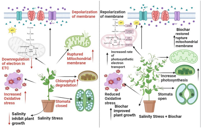
A schematic diagram represents two different mechanisms of pea under salinity stress (control and 80 mM NaCl) with Biochar (8 and 16 g/kg soil).
Introduction
Pea are a significant grain legume that have been used for a long time as animal feed and human food. They are the 4th largely produced beans all around the globe afterwards dry beans, peanuts, and soybeans 1 . The Near East and Mediterranean regions are its primary places of origin 2 . Grown worldwide, it is a cool season, and self-pollinated crop 3 . Peas have potential medical benefits because they comprise of a variety of phytochemicals, such as lectins, saponins, and iso-flavonoids, which are utilized as an anti-cancer agents 4 .
People have cultivated pea ( Pisum sativum L.) from several years, a well-known legume, in various regions of the world. Pea seeds are a good source of protein, fibre, carbs, vitamins, and minerals. Pea seed’s protein has an amino acid profile that is well-balanced and is simple to digest 5 . Pea are planted for their fresh green seeds, soft green pods, dried seeds, and leaves in temperate regions of the world, and they are also grown as a winter crop in subtropics 6 . Compared to many other broad-leaved crops, the pea plant grows more quickly and requires less water 7 . It has been found that exposure to mild salinity (100 mM NaCl) reduces the pea production by 50% and its output is significantly decreased under high saline conditions 8 .
Abiotic factors such as extreme temperatures, salinity, salinity, UV radiation, wounding, or heavy metals are among the many variables that plants must cope with. Additionally, biotic factors are caused by pathogenic bacteria, nematodes, insects, or herbivores 9 . Salinity is one of these abiotic variables that poses a serious risk to agriculture, water resources, and land productivity in coastal regions as well as arid and semi-arid parts of the world 10 . Salinity is not a current problem rather it is an old problem with irrigated agricultural lands, which is more extensively increased by urbanisation, industrialization, and agricultural modernization 11 .
Salinity is a major threat to agricultural land worldwide, diminishing agricultural production and biodiversity, damaging the environment, contaminating groundwater, raising the possibility of flooding, creating issues with food security, and limiting economic progress 12 . According to reports, 450 million hectares of soil in Pakistan 13 and 900 million hectares worldwide 14 are damaged by salinity. By adding additional challenges such as nutritional imbalance, water stress and cytotoxicity caused by increased excretion of sodium (Na + ) and chloride (Cl − ) ions, salt stress inhibits plant growth. Excessive concentrations of Na + and Cl − ions lead to production of reactive oxygen species, which causes oxidative stress in plants 15 .
Excessive salinity accumulation in the rhizosphere of a plant can have detrimental effects on several plant health aspects, such as physiological processes, mineral ion accumulation, damage to PSII reaction centres, and disruption of metabolic processes, which is mainly caused by the production of reactive oxygen species (ROS). As a result, the plant experiences growth retardation and severe impairment of its metabolic processes 16 . Remarkably, many plants try to keep a subtle balance between enzymatic and non-enzymatic antioxidant defence systems in response to severe salt stress 17 . Furthermore, ROS produced by salinity stress might harm molecular structure of plants 18 . Excessive formation of ROS causes the breakdown of chlorophyll to occur more quickly and decreases the photochemical capacity of antioxidants. This can have an impact on lipids, proteins, and nucleic acids, leading to lipid peroxidation, protein degradation, and DNA changes 19 .
To mitigate the noxious impacts of salt stress, several approaches were employed, including the administration of organic and inorganic fertilisers, seed priming, exogenous phytohormone application, and screening of different cultivars 20 , 21 . Biochar has been shown to be a highly effective strategy for increasing crop yield and resilience to abiotic challenges 22 , 23 . Biochar is a carbon-rich substance that is used as a key soil conditioner to improve plant health, soil quality, and resistance to salt stress 24 . An appealing remedy and sustainable technique to repair degraded soil resources is the application of biochar (organic fertilizer) to lessen the adverse effects of salt stress 25 .
Species distributions and associations between soil organisms are greatly influenced by climate change 26 . With due to scenario of surprisingly changing climatic conditions, interactions among various community members are highly impacted that could be advantageous, pathogenic, and have little to no functional impact 27 . Crop rotation has a more significant effect on the community of soil microorganisms. The use of pea, lentil, and chickpea in rotation has an impact on enhancing the activity of arbuscular mycorrhizal fungi associated with wheat 28 . Many studies have also demonstrated that wheat-based crop rotations pose remarkable beneficial impacts on the community of soil microorganisms 29 . Biochar is an environmentally friendly bio stimulant and soil conditioner, primarily boosts agricultural yield while mitigating the negative impacts of various abiotic stressors. Application of biochar to improve soil health, possesses high potential to improve plant growth and yield as well as escalate resistance to abiotic stresses due to its ability to regulate ionic homeostasis, antioxidant machinery, heavy metal accumulations, and oxidative damages 30 . Furthermore, biochar can maintain photosynthetic activity, improve transpiration, protein synthesis, and nutritional uptake, and improve stress tolerance by regulating ROS generation 31 , 32 .
To enhance crop production, biochar may often lessen the negative impacts of climate disruptions (drought, waterlogging, salinity), as well as degraded soils. Additionally, by immobilizing inorganic and/or organic pollutants by surface complexation, electrostatic attraction, ion exchange, adsorption, and co-precipitation, it might lessen the bioavailability and phytotoxicity of pollutants in soils with contamination 33 .
Biochar is produced via low-oxygen pyrolysis of organic materials. There are numerous characteristics of biochar, including a wide surface area, high porosity, and cation exchange capacity, and mineral enrichment 34 , 35 . Biochar is well known for its ability to function as a significant growth regulator, a bio stimulant of agricultural production, and an enhancer of plant development under salt stress 36 . The application of biochar to saline soils has the potential to greatly lessen oxidative stress and promote plant development. As a result, it can be applied as a remedy to lessen the effects of salt stress on agricultural soils. By increasing the potential of antioxidant activities and increasing concentrations of unsaturated fatty acids, biochar application helps plants by supporting numerous processes that improve membrane integrity and plant-water connections. These mechanisms, in turn, lower sodium ion concentrations and MDA (malondialdehyde) levels 37 . Application of biochar has proven to be a more effective strategy for increasing the production of proline, glycine betaine, flavonoids, osmolytes, and glycine 38 . Much research has been conducting nowadays to explore the potential role of organic amendments in soil to alleviate abiotic stresses. Which is gaining more researchers attention due to its cost effectiveness strategy for sustainable agriculture. The core objectives of this study were to evaluate the impact of biochar on growth and physiological attributes of pea varieties under saline conditions, and to investigate the antioxidants, osmolytes and mineral ions activity in pea varieties under salinity stress. This is hypothesized that biochar application in soil can reduce the detrimental effects of salt stress on pea plants.
Methodology
Experimental setup.
A pot experiment on two pea varieties (green grass and meteor) was carried out in the University of Agriculture, Faisalabad in an old botanical garden. Seeds of pea varieties (Meteor and Green Grass) were attained from Ayub Agricultural Research Institute, Faisalabad. The treatments of this study were, (a) Pea varieties; (i) V1 = Meteor and V2 = Green Grass, Stress, (b) Control (0 mM) and (ii) Salinity (80 mM) (c) Biochar applications; (i) Control, (ii) 8 g/kg soil (56 g) and (iii) 16 g/kg soil (112 g). Thirty-six pots were arranged in two groups and filled with 7 kg of soil. In 18 pots seeds of Meteor variety were cultivated and similarly in other 18 pots, seeds of Green Grass variety were sown (nine seeds per pot). The experiment was conducted under a completely randomized design (CRD) along with three replications. The pea plants were moderately irrigated with tap water with a gap of one week throughout cultivation. After two weeks of sowing, when germination occurred plants were thinned as 5 plants per pot were left for treatment. The pH of the soil was 8.5 that is alkaline, the Hoagland’s solution was applied for better growth of pea varieties. Hoagland’s nutrient solution was applied twice, the first time 1000 mL per pot and the second time, 500 mL per pot with a gap of two weeks. Two levels of salt stress (Control and 80 mM) were applied to the soil, after germination of two weeks. Rice straw was used as a raw material to make biochar. Following the two-week of stress period, three levels of biochar (Control, 8 g/kg soil, and 16 g/kg soil) were amended to soil medium.
Harvesting and data collection
Biochar treated plants were harvested after two weeks. Fresh plants were collected for determination of morphological parameters, then fresh plants were kept in the oven at 65 °C for 15 days, to get their dry weight and shoot and root ions analysis. The other plants were kept in plastic zipper bags after harvesting and instantly placed in a freezer at − 15 °C to estimate photosynthetic activity, reactive oxygen species, and antioxidant activity in pea varieties.
Morphological parameters
After harvesting plants consisting of root and shoot parts, morphological indices were determined using measurement scale such as root length and shoot length. To determine the fresh weight of root and shoot of instantly harvested plants, an electronic weighing balance was used. The harvested plant samples were then kept in an oven set to at 65 °C temperature for two weeks to determine the dry weight of shoot and root samples.
Photosynthetic indices
The efficiency of the photosynthetic process was estimated using Arnon method 39 . For this purpose, the contents of Chl. a, Chl. b, total Chl., and carotenoids were determined by following the procedure in which 0.1 g of fresh leaf plant samples were cut into small pieces and placed in small plastic jars containing 5 mL of 80% acetone. Then, small plastic jars were placed at room temperature of 25 °C overnight. The next day the aliquot in a cuvette and the reading was noted at 480, 663, and 645 nm using a spectrophotometer (“IRMECO U2020”, Germany).
Oxidant activities in pea varieties
The activity of hydrogen peroxide (H 2 O 2 ) was evaluated using the method described by Velikova 40 . Samples of fresh plant leaves were ground using a pestle and mortar in 3 mL of a solution containing 0.5% trichloroacetic acid to estimate H 2 O 2 content. The ground material was centrifuged. After 15 min of centrifugation, 0.5 mL of sample extract, 0.5 mL of potassium phosphate buffer, and 1 mL of potassium iodide were added into test tubes and vortexed the sample mixtures for one minute. A spectrophotometer (Model: IRMECO U2020, Germany) was used to measure the reading at 390 nm.
Malondialdehyde concentrations were detected by using protocol given by Cakmak and Horst 41 . For this purpose, 0.3 g of fresh plant leaf sample was crushed in 3 mL of 1% w/v tricarboxylic acid (TCA). The ground samples were then centrifuged at 12,000 rpm for 15 min. Afterward, test tubes were taken in which centrifuged plant samples and 1 mL of 0.5% TBA (thiobarbituric acid) in a 20% TCA solution were added. Finally, test tubes were kept in water bath for 15 min at 95 °C, then kept on ice for 15 min. Reading was noted at 532 nm and 600 nm by using a spectrophotometer (Model: IRMECO U2020, Germany).
Enzymatic antioxidants activities
In a pre-cooled pestle and mortar, 250 mg of fresh leaf samples were crushed by adding 5 mL of potassium phosphate buffer to each sample. After being homogenized, the ground constituent was put into an Eppendorf tube and centrifuged for 15 min at 12,000 rpm. The supernatant solution was separated and poured into another Eppendorf tube and stored at a temperature of 15 °C. Utilizing their measurement techniques, CAT, SOD, and POD activities were assessed.
The procedure for measuring catalase (CAT) activity was disclosed by Chance and Maehly in 1955 42 . A cuvette was filled with 1.9 mL of cold potassium phosphate buffer, 1 mL of H 2 O 2 , and 0.1 mL of plant material. Using the spectrophotometer (IRMECO U2020, Germany), the absorbance was measured at 240 nm at intervals of 0, 30, 60, and 90 s.
By using the method outlined by Chance and Maehly 45 , the peroxidase (POD) activity was assessed. The cuvette was filled with 750 µL phosphate buffer, 0.1 mL of guaiacol, 0.1 mL of H 2 O 2 , and 50 µL of plant extract. In the end, readings of plant samples were recorded at 0, 30, 60, and 90-s intervals using a spectrophotometer (IRMECO U2020, Germany) at 470 nm wavelength.
The antioxidant potential of superoxide dismutase was determined using a Spitz and Oberly method 43 . For this purpose, 0.4 of mL distilled water, 250 mL of cold potassium phosphate buffer, 0.1 mL of L-methionine solution, 0.1 mL of Triton X solution, 0.05 mL of Nitroblue tetrazolium (NBT), 0.05 mL of plant extract, and 0.05 mL of riboflavin solution were all put into plastic cuvettes. Afterward, cuvettes were exposed for 15 min to a fluorescent lamp. Without the plant sample, a blank sample was collected. Finally, readings of plant samples were recorded using an ultraviolet–visible spectrophotometer at 560 nm wavelength.
Non-enzymatic antioxidant activities
Strack and Wray's method 44 was used to calculate the anthocyanin content. For this, 2 mL of acidified methanol and 100 mg of fresh pea leaf were put into test tubes. The samples in the test tubes were then incubated for 60 min at 90 °C. Afterwards, final readings of plant samples were recorded using spectrophotometer at 535 nm.
Flavonoid content of the plant samples was assessed by Ribarova and Atanassova 45 approach following the procedure in which 0.1 g of freshly taken pea plant samples were immersed in 5 mL of 80% acetone and placed overnight. After that, 1 mL of the sample was added in to 4 mL of distilled water in a test tube, the mixture was left for 5 min. A subsequent addition of 0.6 mL of 5% NaNO 2 and 0.5 mL of 10% AlCl 3 , 2 mL of 1 M NaOH and 2.4 mL of distilled water was made in to test tube. Afterward, final readings of plant samples were recorded using a spectrophotometer at 510 nm.
Organic osmolytes
Total soluble protein content of the plant samples was measured by Bradford's approach 46 following the protocol in which 250 g of freshly taken pea plant samples were ground in 5 mL of potassium phosphate buffer using pestle and mortar. The resultant homogenous plant extract was then poured into an Eppendorf tube. Afterwards, the obtained homogeneous plant extract was subjected to a centrifugation process at 12,000 rpm for 15 min. After centrifugation, the supernatant was collected in a separate Eppendorf tube. Subsequently, a reaction was carried out by mixing 1 mL of plant extract sample and 5 mL of Bradford reagent in a test tube. Then samples in the test tube were vortexed and the samples' readings were recorded using a spectrophotometer at 595 nm.
Following the procedure developed by Yoshida 47 , the total soluble sugar content was measured. The Fresh leaf samples weighing 100 mg were placed in test tubes along with 10 mL of distilled water. These samples were then incubated in a water bath set at 90 °C for one hour. Following incubation, samples were diluted up to 50 mL by the addition of distilled water. Separate test tubes were prepared, containing 1.5 mL of the diluted plant sample and 5 mL of the Anthrone reagent. These samples were once again submerged in a 90 °C water bath for 20 min. After cooling to room temperature, absorbance was measured at the wavelength of 620 nm using a spectrophotometer.
Glycine betaine (GB)
To assess the glycine betaine contents in plant samples, 0.25 g of fresh plant shoot sample was taken and ground in 5 mL of distilled water. After that, the ground samples were subjected to a centrifugation process at a speed of 12,000 rpm. Following the centrifugation process, a chemical reaction was carried out by mixing 1 mL of plant extract sample and 1 mL of 2N-H 2 SO 4 . Then 0.5 mL from that prepared extract was taken in a separate test tube. Added 0.2 mL KI solution in test tubes and kept in ice for one and half hours. After a time of 90 min, 6 mL di-chloroethane and 2 mL distilled water were put into test tubes. As a result, two distinct layers of upper and lower were formed inside the test tube and the sample was taken from the lower layer and the readings were recorded on a spectrophotometer at 365 nm wavelength.
Ion analysis
To quantify the uptake of inorganic ions Allen’s approach 48 was used. In that context of mineral ions determination, 0.1 g of over-dried plant sample was taken and put into a digestion flask containing 2 mL of sulphuric acid. The digestion flasks were then enclosed with aluminum foil and placed overnight. The very next day, the digestion flasks were transferred to a hot plate set at 200 °C until the colour of the solution became transparent by adding hydrogen peroxide drop by drop. At the end, solution obtained after digestion was subjected to filtration process and the final volume of the solution obtained after filtration was made up to 50 mL by adding distilled water. Na + , K + and Ca 2+ ions were measured by using this dilute digested solution. Reading was noted by using flame-photometer.
Analytical statistics
The layout of this experiment was set up under the Complete randomized design (CRD) with three factors. After research trial completion, obtained data from plant samples was subjected to statistical analysis and graphical representation using Statistix 8.1, R-studio (v4.3.3), Originpro (2022), and Microsoft Excel (Version, 2016) (Microsoft Corporation, Redmond, WA, USA) software’s.
Study protocol must comply with relevant institutional, national, and international guidelines and legislation
The use of plants in the present study complies with international, national and/or institutional guidelines.
Growth parameter analysis
Accumulation of salt (80 mM) in the growth medium remarkably decreased the biomass production of pea varieties (Table 1 ), such root length decreased by (18%, and 29%), while shoot length up to 29% in each variety and their fresh weight reduced by (41, 44%) and (37, 26%) as well as dry weight reduced by (27, 22%) and (38, 30%) in both pea varieties (Table 1 ). The leaf area of both varieties was reduced by 43% in V1 and 28% in V2 under salt stress (80 mM) (Table 1 ). The amendment of biochar improved the production of biomass by enhancing root and shoot length by (40, 44%) and (39, 57%) in V1 and V2, respectively (Table 1 ). Root fresh weight greatly improved by amendment of biochar up to (37, 59%) and fresh weight of shoot by (35, 36%), in both varieties accordingly (Table 1 ). Leaf area was increased by 55 and 44% in V1 and V2, correspondingly. The overall results of growth attributes manifested that V2 (Green grass) performed better than V1 (Meteor). Moreover, the maximum increment was observed when (16 g/kg soil) of biochar was put into the soil (Table 1 ).
Photosynthetic pigments
Under salinity stress conditions, the V1 and V2 exhibited more significant (p < 0.05) behavior for photosynthetic pigments (Table 2 ). Salinity stress considerably reduced the content of chl a up to (16, 25%) chl b (27, 36%), total chl. (23, 32%) and carotenoid content up to (37, 39%) in V1 and V2, respectively (Table 2 ). The amendment of biochar enhanced all these parameters in V1 and V2 as chl a up to (32 and 40%), chl b (57 and 51%), total chl (48 and 47%) as well as carotenoids (43 and 40%) (Table 2 ). Overall findings showed that for all photosynthetic pigments, V2 exceeded V1. Additionally, the highest elevation was noted with a 16 g/kg soil biochar amendment as opposed to an 8 g/kg soil amendment of biochar (Table 2 ).
Oxidants and antioxidants
The production of MDA and H 2 O 2 was significantly (p < 0.05) enhanced when pea plants were exposed to salt stress (Fig. 2 ). In stress situations, V2 showed (44%) greater H 2 O 2 content than V1 (35%) (Fig. 2 ). In comparison to control plants, the MDA concentration of salt-stressed plants rise by 27% in V1 and 62% in V2 (Fig. 2 ). However, the external application of biochar managed to lower the H 2 O 2 level in V1 and V2 to 31% and 23%, respectively (Fig. 2 ). Similarly, the addition of biochar reduced the MDA contents in V1 and V2 up to 26% and 33%, respectively, in comparison to the control (Fig. 2 ). The acquired data indicated that in salt-stressed plants, the activities of SOD, POD, and CAT were elevated in both types (Table 2 ). Under salt stress conditions, there was a maximal increase in activities of SOD (69%) and POD (63%), as well as (196%) increase in CAT activities in V1, in addition, enhancements of 67, 122, and 87% were observed in V2 (Table 2 ). However, the addition of biochar further boosted the antioxidant activities in both V1 and V2 by 37% and 22% for SOD, 48% and 38% for POD, as well as 22% and 27% for CAT, respectively, compared to salt stress (Table 2 ). The activity of Anthocyanin was decreased by 33 and 44% in V1 and V2, while that of flavonoid was enhanced by 46 and 80% in both varieties, under salinity stress (80 mM) (Fig. 2 ). The application of biochar substantially enhanced the anthocyanin content by 58 and 54% in V1 and V2, and further enhanced flavonoid content by 37 and 30% in both pea varieties (Fig. 2 ). Furthermore, compared to biochar (8 g/kg soil), a greater increase was seen with a 16 g/kg biochar amendment (Fig. 2 ).

Effect of salinity and biochar on ( A ) Hydrogen peroxide (µmol g −1 F.Wt), ( B ) Malondialdehyde (µmol g −1 F.Wt), ( C ) Anthocyanin (Units mg −1 F.Wt), ( D ) Flavonoids (µg g −1 F.Wt), of pea. Error bars above means specify the ± SE of three replicates. Same letter sharing by means for a parameter do not vary significantly at p ≤ 0.05. V1 = Meteor; V2 = Green Grass; Control = No salinity; Salinity = 80 mM Nacl; Control = No Biochar; BC (55 g) = Biochar 8 g/kg soil; BC (110 g) = Biochar 16 g/kg soil.
Different organic osmolytes including total soluble proteins, total soluble sugars, and glycine betaine were recorded to analyze the individual and cumulative effects of salt stress and biochar on V1 and V2 (Fig. 3 ) Analysis of variance revealed that the following osmolyte contents increased under salinity stress (80 mM) as, TSP (78, 57%) and GB up to (29, 120%) in both varieties, correspondingly (Fig. 3 ). While, furthermore enhanced by the application of biochar as TSP (26 and 31%), and GB (73 and 67%) in V1 and V2, respectively (Fig. 3 ). Under salinity stress, TSS was reduced by (35 and 31%) while enhanced by 47 and 36% on the application of biochar as compared to the salt-stressed plants (Fig. 3 ). Additionally, 16 g/kg soil, biochar addition showed a major improvement when compared to biochar (8 g/kg soil) (Fig. 3 ).

Effect of salinity and biochar on ( A ) total soluble protein (mg −1 g F.W), ( B ) total soluble protein (mg −1 g F.W), ( C ) and Glycine betaine (µg g −1 g F.Wt) of pea. Error bars above means specify the ± SE of three replicates. Same letter sharing by means for a parameter do not vary significantly at p ≤ 0.05. V1 = Meteor; V2 = Green Grass; Control = No salinity; Salinity = 80 mM Nacl; Control = No Biochar; BC (55 g) = Biochar 8 g/kg soil; BC (110 g) = Biochar 16 g/kg soil.
Inorganic ions
Accumulation of salt through rooting and growth medium remarkably declined the mineral ions as, shoot calcium (52, 30%), root calcium (45, 41%), shoot potassium (28, 23%), root potassium up to (41, 34%), while (80 mM) NaCl enhanced the uptake of shoot sodium by (63 and 71%) as well as root sodium up to (24 and 49%) in V1 and V2, respectively (Fig. 4 ). The application of biochar significantly improved the activity of inorganic ions in both varieties (V1, V2) as, Shoot Ca 2+ (99, 76%), root Ca 2+ (68, 51%), shoot K + up to (48, 29%) and root K + (74, 72%) (Fig. 4 ). On the other hand, the uptake of Na + through root and shoot reduced on application of biochar as, root Na + up to (30, 25%) and shoot Na + decreased up to (22, 31%) (Fig. 4 ). Overall results demonstrated that the uptake of Ca 2+ and K + was enough in V2 than in V1 (Fig. 4 ). Moreover, the greater increase was observed with 16 g/kg soil biochar amendment as compared to biochar (8 g/kg soil) (Fig. 4 ).

Effect of salinity and biochar on ( A ) shoot Ca 2+ (mg −1 g D.W), ( B ) root Ca 2+ (mg −1 g D.W) ( C ) shoot K + (mg −1 g D.W), ( D ) root K + (mg −1 g D.W), ( E ) shoot Na + (mg −1 g D.W), ( F ) and root Na + (mg −1 g D.W), of pea. Error bars above means specify the ± SE of three replicates. Same letter sharing by means for a parameter do not vary significantly at p ≤ 0.05. V1 = Meteor; V2 = Green Grass; Control = No salinity; Salinity = 80 mM Nacl; Control = No Biochar; BC (55 g) = Biochar 8 g/kg soil; BC (110 g) = Biochar 16 g/kg soil.
Correlation analysis
The correlation matrix shows strong positive and strong negative correlations among various parameters of pea varieties under salinity stress (Fig. 5 ). The correlation analysis showed that the growth parameters including shoot and root length, shoot and root fresh weight, shoot and root dry weight, leaf area, chl a , chl b , total chlorophyll, and carotenoids, were negatively correlated with CAT, SOD, POD and H 2 O 2 , MDA, flavonoids, and TSP, shoot and root Na + ions (Fig. 5 ). The morphological parameters including shoot and root fresh and dry weight and their lengths were positively correlated with anthocyanin, TSS, shoot and root K + and Ca 2+ ions, respectively. In addition, the correlation was strongly positive among morphological and anthocyanin, total soluble sugars, and potassium and calcium ions (Fig. 5 ).

Correlation matrix between morpho-physiological, biochemical attributes and ionic contents of pea (Meteor and Green Grass) varieties.
Heatmap analysis
A two-way heatmap with a dendrogram was drawn to observe the role of biochar on various observations of pea under salinity stress conditions (Fig. 6 ). The observations were divided into groups according to how similar they were during different treatment phases, and the relationships between the groups were shown by coloured squares. The colour (Navy blue) exhibited a strong positive association while the colour (Maroon) exhibited a strong negative correlation for various observations, impacted by biochar under salinity stress (Fig. 6 ). Heatmap has clustered into four groups. In the first group, TSP, SOD, flavonoids, and CAT were clustered. These parameters are strongly positively correlated with salinity stress (80 mM) and biochar (112 g) and weakly correlated, at biochar (56 g) under salinity (80 mM) stress. Under (0 mM) and biochar (56 g) conditions, above-mentioned attributes, showed weak correlation while strongly negatively correlated under (0 mM) and control (No biochar) conditions, respectively. This group demonstrated that the application of biochar (112 g) improved the levels of organic osmolytes and antioxidants that mitigated the adverse effects of oxidative damage caused by salinity stress. The second group included GB and POD that were strongly positively correlated at salinity (80 mM) and biochar (112 g) while weakly correlated at salinity (80 mM) and biochar (56 g) while strong negative correlated at control (0 mM) and control (No biochar) and weakly negative correlated at control (0 mM) and biochar (56 g). These observations showed that under salinity stress GB and POD activity of pea varieties improved by application of biochar. The third group contained (Shoot and root Na + , MDA, and H 2 O 2 ). These attributes were strong positively correlated at (80 mM) and control (No biochar) while showed negative correlation at salinity (0 mM) and biochar (112 g). These findings showed that the application of NaCl (80 mM) increased the uptake of Na + ions in the shoot and root as well as H 2 O 2 and MDA of pea plants. In fourth group leaf area, shoot and root fresh and dry weight, their lengths Ca 2+ and K + ions and photosynthetic pigments (chl a , b , total chlorophyll, and carotenoids) and anthocyanin were clustered. These parameters were strongly positively correlated at control (0 mM) and biochar (112 g), and weakly correlated at salinity (0 mM) and biochar (56 g) while strong negatively correlated at salinity (80 mM) and control (No biochar) and weakly negative correlated at NaCl (80 mM) and biochar (56 g). These interpretations showed that under salinity stress (80 mM), growth attributes and photosynthetic pigments of pea varieties improved by the application of biochar. (Fig. 6 ).

Heatmap with dendrogram between morpho-physiological, biochemical attributes and ionic contents of pea (Meteor and Green Grass) varieties.
Around the world, one of the major problems that many agricultural regions face is soil salinity. Salt stress prevents the effective growth of plants in addition to having an impact on germination. Pea plant growth, physiological, and biochemical attributes were shown to have significantly decreased as a result of salt stress during the current study of biochar's potential in response to saline stress. According to earlier studies, biochar plays a major contribution in enhancing plant stress tolerance 49 , 50 .
Applying biochar to soils has been proposed to be an environmentally friendly organic additive to improve soil health and quality, enhance moisture-holding capacity, boost cation exchange capacity, increase organic matter status, and increase soil fertility by retaining nutrients and encouraging microbial activity 51 . The deleterious impact of salt stress may be the cause of the decrease in plant height and physiological attributes (Fig. 1 ). Mainly one key abiotic factor that restricts agricultural productivity and growth is salinity 52 . Salinity may harm plants because it lowers the water potential of the soil, which can lead to osmotic stress, a decrease in water availability, and a slowdown in plant growth 53 . These findings are from research conducted by Elsakhawy 54 and El-Banna and Abdelaal 55 . The application of biochar improved plant growth indices, reduced plant signs of salinity stress, and increased the growth of the plants 56 . The aforementioned growth features significantly increased after being treated with biochar 50 , 58 . The positive effects of biochar might stem from its ability to improve the chemical and physical characteristics of soil by lowering the pH and sodium concentration in salinized soil 59 . Conversa 60 demonstrated that the biochar's favorable outcomes might be attributed to the emergence of a more advantageous microbial population as well as the availability of potassium and phosphorus. Salinity concentrations between 1500 and 3000 ppm in our experiment caused a decrease in the area of leaves per plant in pea (Fig. 2 ). These results may be attributed to salinity's detrimental effects on water uptake, which results in less water flowing from the root to the leaves and less cell division, which in turn reduces the area of leaves per plant 61 . Previous studies also demonstrated that there is great potential for using biochar to improve K + uptake and reduce Na + uptake. The use of biochar promoted the growth and size of root cells, enabled rice roots to absorb more K + , and prevented ion toxicity by keeping Na + ions out of the xylem and isolating them in the vacuole when exposed to salt stress 62 .
Chlorophyll a and b contents in pea plants were significantly reduced when they were exposed to salinity (Fig. 3 ); this outcome was consistent with findings from Elsaeed 63 and Zamin 64 . El-Esawi 65 suggest that salinity may have a negative impact on photosynthetic rate, CO 2 assimilation, stomatal movement, and the production of the enzyme chlorophyllase. Severe atmospheric conditions that are detrimental for photosynthetic equipment include salt, water stress, temperature, and heavy metals. The salt-induced decrease in chlorophyll concentration reported in this work may be explained by increased chlorophyllase activity, which breaks down pigment proteins and ultimately lowers chlorophyll content in plants. Salinity-induced chlorophyll reduction may be primarily caused by changes in the chlorophyll complexes of proteins 66 , the disintegration of the chlorophyll via free radical damage-induced ROS, the deterioration of the thylakoid membrane, and decreases in the formation of chlorophyll or accelerated enzymatic destruction of chlorophyll 67 . Moreover, it has been demonstrated that salinity lowers the concentration of intermediate substances in the biosynthesis of chlorophyll and regulates the expression of many genes encoding the Mg-chelatase subunit 68 . Treatments with biochar improved the number of leaves, the concentration of chlorophyll a and b, and RWC in salt-stressed pea plants. Similar to the findings of Haider 69 , the critical role that biochar plays in enhancing prior features is linked to enhanced water and nutrient uptake, water-holding capacity, and the significant avoidance of chlorophyll degradation under salt stress. Improving stomatal density and increasing water availability while reducing ROS buildup 70 . With more chlorophyll a and b contents production as a result of biochar's superior coupling effects on salt-stressed pea plants, photosynthesis the most significant physiological process in all plants increased, improving enzyme activity under salinity stress.
MDA and ROS, primarily SOD and H 2 O 2 , were significantly elevated under salinity conditions with the two salinity levels (Fig. 4 ). Under different stresses, the same outcomes were noted 71 . According to Ahanger 72 and Kamran 73 , salinity may hurt metabolic dysfunction, nutritional imbalance, membrane stability, and oxidative stress. Under salt stress, these results are consistent with those of Hasan 74 . On the other hand, when biochar was added to stressed plants, MDA and ROS levels decreased. Biochar may have these positive effects because it improves soil structure, increases water circulation, and increases pea production 75 . Thus, in addition to lowering ROS, reducing oxidative damage, and improving plant growth and development of stressed plants were the outcomes of improving enzymatic and nonenzymatic antioxidant systems. According to our findings, the stressed plants have significantly higher levels of CAT, POX, and SOD (Fig. 5 ). Because salinity causes oxidative stress and raises MDA and ROS, it may also have an impact on enzyme activities 76 . Increased CAT, POD, and SOD enzyme activity was necessary to maintain osmotic potential and is essential for ROS scavenging. Treatments with biochar decreased the buildup of Na + and Cl − , which in turn decreased the concentration of ions in the stressed plants and ultimately increased the activity of the enzymes 77 . To remove the excess ROS production, the plants attempted to activate their defensive system (CAT, POD, and SOD) in response to the salinity 78 .
According to Waqas 79 , biochar stimulates hormone production in plants, giving rise to a strong defence against various stressors. Through several processes, the addition of biochar to the soil improved the overall performance of the plants (Fig. 6 ). According to Jaiswal 80 and Wang 81 , they improved photosynthesis and carbon fixation, reduce chlorophyll degradation, and regulate the homeostasis of minerals in the soil. Our study's findings also demonstrated that pea seedlings accumulated the osmoregulatory components glycine betaine (GB) and proline in response to salinity stress. According to Paradisone 82 , GB has been shown to serve a variety of roles as a compatible solute in osmotic adjustment under salt stress. These roles include stabilizing enzyme and protein structures, protecting both protein and membrane functions from harmful amounts of Na + and Cl − ions, and eliminating excess ROS. Moreover, GB may indirectly cause modifications in coenzyme turnover efficacy, which is necessary to sustain respiration rates and photosynthesis during stressful situations 83 .
The findings of our study showed that salt stress caused pea plants to have higher Na + and lower K + and Ca 2+ contents. The current results were supported by earlier research. Harmful sodium ion blocks the absorption of water and vital nutrients during high salinity exposure 84 , which causes osmotic stress and water loss in cells 85 . Potassium ion (K + ) uptake is restricted by the buildup of Na + in the cells of plants, which is necessary for the growth of plants 86 (Nguyen et al., 2021). Excessive salt levels caused a decrease in K + and Ca 2+ uptake but a rise in Na + absorption 87 . According to Okhovatian-Ardakani 88 , excessive Na + inflow disrupts ion homeostasis, causing sudden changes in enzyme activity and oxidative damage. The integrity and proper operation of cellular membranes depend on balanced concentrations of K + and Ca 2+ ions 89 . Plant resistance to salt involves preventing Na + influx, improving K + absorption, and/or maintaining K + homeostasis 90 . Under salt stress, salt-tolerant cultivars keep high concentrations of K + and Ca 2+ ions 91 . Calcium (Ca 2+ ) works as a molecule that transfers signals and plays a key role in regulating ionic balance or osmotic balance 92 . Plants can produce high-yield crops by increasing their photosynthetic rate through the buildup of inexpensive osmoticum 93 . Salt stress is emerging as a concerning factor for plant growth and development among many stressors 94 , 95 , 96 .
In this investigation, biochar applied topically greatly raised the K + and Ca 2+ concentrations in the shoots and roots of both pea cultivars. The explanation that was given was that the presence of BC in the soil facilitates the adsorption and availability of nutrients as well as the leaching of macronutrients from the root zone, which may be the primary cause of spinach's higher nutritional levels 97 . The potential of biochar application in terms of increasing the availability of potassium contents in the soil and providing more pivotal nutrients proved to be a more strategic practice in the improvement of pea plants growth under saline conditions 98 . Because of its porous structure, large surface area, negative surface charge, and ability to increase the soil's cation exchange capacity, the use of biochar improved soil nutrient cycling, including N, P, and K for plant intake. This allowed nutrient sequestration, which increased plant N, P, and K concentrations under water deficit and soil salinity conditions 99 .
Salinity stress reduced the morpho-physiological and biochemical attributes of pea verities leading to reduced nutrient uptake by the plants. However, biochar improved the plant growth attributes by improving the photosynthetic rate and water status of plants. In addition, biochar improved the activities of enzymatic and non-enzymatic antioxidants such as CAT, SOD, POD, TSP, and TSS, and maximum improvement was observed at 16 g/kg soil biochar. In short, the present study suggested that biochar (16 g/kg soil) may help to mitigate salinity stress adversities in pea through maintaining photosynthetic pigments and improved ionic attributes. Furthermore, the analysis at molecular levels needs to be studied in pea under salinity-stressed conditions.
Data availability
All data generated or analyzed during this study are included in this published article.
Farshi, P., Mirmohammadali, S. N., Rajpurohit, B., Smith, J. S. & Li, Y. Pea protein and starch: Functional properties and applications in edible films. J. Agric. Food Res. 15 , 100927 (2023).
Smýkal, P., Coyne, C., Redden, R. & Maxted, N. Peas. Genetic and Genomic Resources of Grain Legume Improvement 41–80 (Elsevier, 2023).
Kyzek, S. et al. Cold atmospheric pressure plasma can induce adaptive response in pea seeds. Plasma Chem. Plasma Process. 39 , 475–486 (2019).
Article CAS Google Scholar
Rungruangmaitree, R. & Jiraungkoorskul, W. Pea, Pisum sativum, and its anticancer activity. Pharmacogn. Rev. 11 (21), 39 (2017).
Article CAS PubMed PubMed Central Google Scholar
Kumari, T. & Deka, S. C. Potential health benefits of garden pea seeds and pods: A review. Legume Sci. 3 (2), e82 (2021).
Rana, C. et al . Stability analysis of garden pea ( Pisum sativum L.) genotypes under North Western Himalayas using joint regression analysis and GGE biplots. Genetic Resour. Crop Evol. 68 , 999–1010 (2021).
Benjamin, J. G. & Nielsen, D. C. Water deficit effects on root distribution of soybean, field pea and chickpea. Field Crop Res. 97 (2–3), 248–253 (2006).
Article Google Scholar
Shahid, S. et al. Proline-induced modifications in morpho-physiological, biochemical and yield attributes of pea ( Pisum sativum L.) cultivars under salt stress. Sustainability 14 (20), 13579 (2022).
Ahmed, S. R. et al. Potential role of silicon in plants against biotic and abiotic stresses. Silicon 15 (7), 3283–3303 (2023).
Ali, Q. et al. Synergistic effects of Rhizobacteria and salicylic acid on maize salt-stress tolerance. Plants 12 (13), 2519 (2023).
Ali, Q. et al. Comparative efficacy of different salt tolerant rhizobial inoculants in improving growth and productivity of Vigna radiata L. under salt stress. Sci. Rep. 13 (1), 17442 (2023).
Abbas, A. et al . Investigating the dynamic responses of Aegilops tauschii Coss. to salinity, drought, and nitrogen stress: A comprehensive study of competitive growth and biochemical and molecular pathways. Front. Plant Sci. 14 , 1238704 (2023).
Syed, A., Sarwar, G., Shah, S. H. & Muhammad, S. Soil salinity research in 21st century in Pakistan: Its impact on availability of plant nutrients, growth and yield of crops. Commun. Soil Sci. Plant Anal. 52 (3), 183–200 (2021).
Hernández-Herrera, R. M. et al. Seaweed extract improves growth and productivity of tomato plants under salinity stress. Agronomy 12 (10), 2495 (2022).
Rodríguez Coca, L. I. et al . Effects of sodium salinity on rice ( Oryza sativa L.) cultivation: A review. Sustainability 15 (3), 1804 (2023).
Farhat, F. et al . Exogenous application of moringa leaf extract confers salinity tolerance in sunflower by concerted regulation of antioxidants and secondary metabolites. J. Soil Sci. Plant Nutr. 23 , 1–17 (2023).
Shafiq, F., Iqbal, M., Ashraf, M. A. & Ali, M. Foliar applied fullerol differentially improves salt tolerance in wheat through ion compartmentalization, osmotic adjustments and regulation of enzymatic antioxidants. Physiol. Mol. Biol. Plants 26 , 475–487 (2020).
Saqib, M., Abbas, G. & Akhtar, J. Root-mediated acidification and resistance to low calcium improve wheat ( Triticum aestivum ) performance in saline-sodic conditions. Plant Physiol. Biochem. 156 , 201–208 (2020).
Article CAS PubMed Google Scholar
Kohli, S. K., Khanna, K., Bhardwaj, R., Abdallah, E. F., Ahmad, P. & Corpas, F. J. Assessment of subcellular ROS and NO metabolism in higher plants: Multifunctional signaling molecules. Antioxidants 8 (12), 641 (2019).
Rhaman, M. S. et al. Seed priming with phytohormones: An effective approach for the mitigation of abiotic stress. Plants 10 (1), 37 (2020).
Article PubMed PubMed Central Google Scholar
Abdel Latef, A. A. H., Tahjib-Ul-Arif, M. & Rhaman, M. S. Exogenous auxin-mediated salt stress alleviation in faba bean ( Vicia faba L.). Agronomy 11 (3), 547 (2021).
Haider, F. U. et al . Biochar application for remediation of organic toxic pollutants in contaminated soils; An update. Ecotoxicology and Environmental Safety 248 , 114322 (2022).
CAS Google Scholar
Ali, M.F. et al. Response of sorghum and millet to poultry and farmyard manure–based biochar treatments. Arabian Journal of Geosciences 15 , 1592 (2022).
Ali, E. F., Al-Yasi, H. M., Kheir, A. M. & Eissa, M. A. Effect of biochar on CO 2 sequestration and productivity of pearl millet plants grown in saline sodic soils. J. Soil Sci. Plant Nutr. 21 , 897–907 (2021).
Ran, C. et al. Benefits of biochar for improving ion contents, cell membrane permeability, leaf water status and yield of rice under saline–sodic paddy field condition. J. Plant Growth Regul. 39 (1), 370–377 (2020).
van der Putten, W. H. Climate change, aboveground-belowground interactions and species range shifts. Annu. Rev. Ecol. Evol. Syst. 43 , 365–383 (2012).
Vandenkoornhuyse, P., Quaiser, A., Duhamel, M., Le Van, A. & Dufresne, A. The importance of the microbiome of the plant holobiont. New Phytologist 206 (4), 1996–1206 (2015).
Borrell, A. N. et al. Fungal diversity associated with pulses and its influence on the subsequent wheat crop in the Canadian prairies. Plant Soil 414 , 13–31 (2017).
Woo, S. L. et al. Pea-wheat rotation affects soil microbiota diversity, community structure, and soilborne pathogens. Microorganisms 10 (2), 370 (2022).
Ali, A. et al . Mitigating ammonia and greenhouse gaseous emission from arable land by co-application of zeolite and biochar. Frontiers in Plant Science 13 , 950944 (2022).
Yasin, M.U. et al . The synergistic potential of biochar and nanoparticles in phytoremediation and enhancing cadmium tolerance in plants. Chemosphere 354 , 141672 (2024).
Murtaza, G. et al . Recent trends and economic significance of modified/functionalized biochars for remediation of environmental pollutants. Scientific Reports 14 , 217 (2024).
Article ADS CAS PubMed PubMed Central Google Scholar
Hasnain, M. et al. Biochar-plant interaction and detoxification strategies under abiotic stresses for achieving agricultural resilience: A critical review. Ecotoxicol. Environ. Saf. 249 , 114408 (2023).
Singh, B. P., Cowie, A. L. & Smernik, R. J. Biochar carbon stability in a clayey soil as a function of feedstock and pyrolysis temperature. Environ. Sci. Technol. 46 (21), 11770–11778 (2012).
Article ADS CAS PubMed Google Scholar
Lehmann, J., Kuzyakov, Y., Pan, G. & Ok, Y. S. Biochars and the plant-soil interface. Plant Soil 395 , 1–5 (2015).
Haider, I. et al . Potential effects of biochar application on mitigating the drought stress implications on wheat ( Triticum aestivum L.) under various growth stages. J. Saudi Chem. Soc. 24 (12), 974–981 (2020).
Ndiate, N. I. et al . Co-application of biochar and arbuscular mycorrhizal fungi improves salinity tolerance, growth and lipid metabolism of maize ( Zea mays L.) in an alkaline soil. Plants 10 (11), 2490 (2021).
Soliman, M. H. et al. Biochar and selenium nanoparticles induce water transporter genes for sustaining carbon assimilation and grain production in salt-stressed wheat. J. Plant Growth Regul. 42 (3), 1522–1543 (2023).
Arnon, D. T. Copper enzymes in isolated chloroplasts polyphenaloxidase in Beta vulgaris. Plant Physiol. 24 , 1–15 (1949).
Valikova, V., Yordano, I. & Edreva, A. Oxidative stress and some antioxidant systems in acid rain-treated bean plants: Protective role of endogenous polyamines. Plant Sci. 151 , 59–66 (2000).
Camak, I. & Host, J. H. Effects of aluminum on lipid peroxidation, superoxide dismutase, catalase and peroxidase activities on root tips of soybean ( Glycine max ). Physiol. Plant. 83 , 463–468 (1991).
Chance, B. & Mahelay, A. Assay of catalase and peroxidase. Methods Enzymol. 2 , 764–817 (1955).
Spitz, D. R. & Oberly, L. W. Measurement of MnSOD and CuZnSOD activity in mammalian tissue homogenates. Curr. Protoc. Toxicol. 8 , 751–758 (2001).
Strack, D. & Wray, V. Anthocyanins. Methods Plant Biochem. 1 , 325–356 (1989).
Ribarova, F. & Atanassova, M. Total phenolics and flavonoids in Bulgarian fruits and vegetables. J.U. Chem. Metal. 40 , 255–260 (2005).
Google Scholar
Bradford, M. M. A rapid and sensitive method for the quantification of microgram quantities of protein utilizing the principle of dye-binding. Ann. Biochem. 72 , 248–254 (1976).
Yoshida, S., Forno, D., Cock, J. & Gomez, K. Determination of sugars and starch in plant tissue. Laboratory manual for physiological studies of rice. Int. Rice Res. Inst. 3 , 46–49 (1976).
Allen, S. E., Grimshaw, M. H. & Rowland, A. P. Chemical analysis. In Methods in Plant Ecology 2nd edn (eds Moore, P. D. & Chapman, S. B.) 234–258 (Springer, 1986).
El-Esawi, et al . Genetic variation and alleviation of salinity stress in barley ( Hordeum vulgare L.). Molecules 23 (10), 2488 (2018).
Rehman, M. et al. Influence of rice straw biochar on growth, antioxidant capacity and copper uptake in ramie ( Boehmeria nivea L.) grown as forage in aged copper-contaminated soil. Plant Physiol. Biochem. 138 , 121–129 (2019).
Hafez, E. M. et al. Differences in physiological and biochemical attributes of wheat in response to single and combined salicylic acid and biochar subjected to limited water irrigation in saline sodic soil. Plants 9 , 1346 (2020).
Kanwal, R. et al . Exogenous ascorbic acid as a potent regulator of antioxidants, osmo-protectants, and lipid peroxidation in pea under salt stress. BMC Plant Biology 24 , 247 (2024).
Farooqi, Z.U.R. et al . Assessment of the change in soil properties and aggregates formation of freshly restored texturally different marginally salt-affected soils under various soil amelioration strategies. Communications in Soil Science and Plant Analysis 61 , 714-722 (2024).
Elsakhawy, T. et al. Efficacy of mushroom metabolites ( Pleurotus ostreatus ) as a natural product for the suppression of broomrape growth (Orobanche crenata Forsk) in faba bean plants. Plants 9 (10), 1265 (2020).
El-Banna, M. & Abdelaal, K. A. Response of strawberry plants grown in the hydroponic system to pretreatment with H2O2 before exposure to salinity stress. J. Plant Prod. 9 , 989–1001 (2018).
Acosta-Motos, J. R. et al. Plant responses to salt stress: Adaptive mechanisms. Agronomy 7 (1), 18 (2017).
El-Naggar, A. et al. Biochar influences soil carbon pools and facilitates interactions with soil: A field investigation. Land Degrad. Dev. 29 (7), 2162–2171 (2018).
Torabian, S., Zahedi, M. & Khoshgoftarmanesh, A. Effect of foliar spray of zinc oxide on some antiox idant enzymes activity of sunflower under salt stress (2018).
Conversa, G., Bonasia, A., Lazzizera, C. & Elia, A. Influence of biochar, mycorrhizal inoculation, and fertilizer rate on growth and flowering of Pelargonium ( Pelargonium zonale L.) plants. Front. Plant Sci. 6 , 429 (2015).
Hafez, Y. et al. Alleviating the detrimental impacts of salt stress on morpho-physiological and yield characters of rice plants ( Oryza sativa L.) using actosol, Nano-Zn and Nano-Si. Fresenius Environ. Bull. 29 (8), 6882–6897 (2020).
Zhang, Y., Ding, J., Wang, H., Su, L. & Zhao, C. Biochar addition alleviate the negative effects of drought and salinity stress on soybean productivity and water use efficiency. BMC Plant Biol. 20 , 288 (2020).
Elsaeed, S. M. et al. Biochar grafted on CMC-terpolymer by green microwave route for sustainable agriculture. Agriculture 11 (4), 350 (2021).
Zamin, M. et al. Developing the first halophytic turfgrasses for the urban landscape from native Arabian desert grass. Environ. Sci. Pollut. Res. 27 , 39702–39716 (2020).
El-Esawi, M. A. et al. Genetic variation and alleviation of salinity stress in barley ( Hordeum vulgare L.). Molecules 23 (10), 2488 (2018).
Siddiqui, M. H. et al. Exogenous melatonin counteracts NaCl-induced damage by regulating the antioxidant system, proline and carbohydrates metabolism in tomato seedlings. Int. J. Mol. Sci. 20 , 1–23 (2019).
Mohamed, H. I., Akladious, S. A. & El-Beltagi, H. S. Mitigation the harmful effect of salt stress on physiological, biochemical and anatomical traits by foliar spray with trehalose on wheat cultivars. Fresenius Environ. Bull. 26 , 4082–4087 (2017).
Szafrańska, K., Reiter, R. J. & Posmyk, M. M. Melatonin improves the photosynthetic apparatus in pea leaves stressed by paraquat via chlorophyll breakdown regulation and its accelerated de novo synthesis. Front. Plant Sci. 8 , 1–10 (2017).
Haider, G., Steffens, D., Moser, G., Müller, C. & Kammann, C. I. Biochar reduced nitrate leaching and improved soil moisture content without yield improvements in a four-year field study. Agric. Ecosyst. Environ. 237 , 80–94 (2017).
Abbas, T. et al. Effect of biochar on cadmium bioavailability and uptake in wheat ( Triticum aestivum L.) grown in a soil with aged contamination. Ecotoxicol. Environ. Saf. 140 , 37–47 (2017).
Esmail, S. M., Omara, R. I., Abdelaal, K. A. & Hafez, Y. M. Histological and biochemical aspects of compatible and incompatible wheat-Puccinia striiformis interactions. Physiol. Mol. Plant Pathol. 106 , 120–128 (2019).
Ahanger, M. A., Tomar, N. S., Tittal, M., Argal, S. & Agarwal, R. Plant growth under water/salt stress: ROS production; antioxidants and significance of added potassium under such conditions. Physiol. Mol. Biol. Plants 23 , 731–744 (2017).
Kamran, M. et al. An overview of hazardous impacts of soil salinity in crops, tolerance mechanisms, and amelioration through selenium supplementation. Int. J. Mol. Sci. 21 (1), 148 (2019).
Hasan, M. M., Baque, M. A., Habib, M. A., Yeasmin, M. & Hakim, M. A. Screening of salt tolerance capability of wheat genotypes under salt stress condition. Univ. J. Agric. Res. 5 (4), 235–249 (2017).
Tammeorg, P. et al. Biochar application to a fertile sandy clay loam in boreal conditions: Effects on soil properties and yield formation of wheat, turnip rape and faba bean. Plant Soil 374 , 89–107 (2014).
Elkelish, A. A., et al. Calcium availability regulates antioxidant system, physio-biochemical activities and alleviates salinity stress mediated oxidative damage in soybean seedlings. J. Appl. Bot. Food Qual. 92 (2019).
Ahmad, P. et al. Jasmonates: Multifunctional roles in stress tolerance. Front. Plant Sci. 7 , 813 (2016).
Fan, H. F., Ding, L., Xu, Y. L. & Du, C. X. Antioxidant system and photosynthetic characteristics responses to short-term PEG-induced drought stress in cucumber seedling leaves. Russ. J. Plant Physiol. 64 , 162–173 (2017).
Waqas, M. A. et al. Potential mechanisms of abiotic stress tolerance in crop plants induced by thiourea. Front. Plant Sci. 10 , 1336 (2019).
Jaiswal, A. K. et al. Molecular insights into biochar-mediated plant growth promotion and systemic resistance in tomato against Fusarium crown and root rot disease. Sci. Rep. 10 (1), 13934 (2020).
Wang, X. et al. Photosynthetic carbon fixation and sucrose metabolism supplemented by weighted gene co-expression network analysis in response to water stress in rice with overlapping growth stages. Front. Plant Sci. 13 , 864605 (2022).
Paradisone, V. et al. Roles of some nitrogenous compounds protectors in the resistance to zinc toxicity in Lactuca sativa cv. Phillipus and Brassica oleracea cv. Bronco Acta Physiol. Plant 37 , 1–8 (2015).
De la Torre-González, A., Montesinos-pereira, D., Blasco, B. & Ruiz, J. M. Influence of the proline metabolism and glycine betaine on tolerance to salt stress in tomato ( Solanum lycopersicum L.) commercial genotypes. J. Plant Physiol. 231 , 329–336 (2018).
Article PubMed Google Scholar
Carpici, E. B., Celik, N. & Bayram, G. The effects of salt stress on the growth, biochemical parameter and mineral element content of some Maize ( Zea mays L.) cultivars. Afr. J. Biotechnol. 9 , 6937–6942 (2010).
Tariq, A. et al . Proteomic and genomic approaches for antioxidant enzyme-mediated defense analyses in higher plants. In Antioxidant Defense in Plants 57–70 (Springer, 2022.)
Nguyen, H. T. T. et al. Rapid accumulation of proline enhances salinity tolerance in Australian wild rice Oryza australiensis domin. Plants 10 , 2044 (2021).
Sultana, A., Mia, M. A. B. & Ahmed, J. U. Cation dynamics and growth morphology of aromatic rice under salt stress. Bangladesh J. Sci. Ind. Res. 56 , 207–214 (2021).
Okhovatian-Ardakani, A. R., Mehrabanian, M., Dehghani, F. & Akbarzadeh, A. Salt tolerance evaluation and relative comparison in cuttings of different omegranate cultivar. Plant Soil Environ. 56 , 176–185 (2010).
Shihab, M. O. & Hamza, J. H. Seed priming of sorghum cultivars by gibberellic and salicylic acids to improve seedling growth under irrigation with saline water. J. Plant Nutr. 43 , 1951–1967 (2020).
Teakle, N. L. et al. Differential tolerance to combined salinity and o 2 deficiency in the halophytic grasses Puccinellia ciliata and Thinopyrum ponticum : The importance of K+ retention in roots. Environ. Exp. Bot. 87 , 69–78 (2013).
Afzal, I., Rauf, S., Basra, S. M. A. & Murtaza, G. Halopriming improves vigor, metabolism of reserves and ionic contents in wheat seedlings under salt stress. Plant Soil Environ. 54 , 382–388 (2008).
Shabbir, R. et al. Calcium homeostasis and potential roles to combat environmental stresses in plants. South Afr. J. Bot. 148 , 683–693 (2022).
Abideen, Z. et al. Moderate salinity stimulates growth and photosynthesis of phragmites karka by water relations and tissue specific ion regulation. Environ. Exp. Bot. 105 , 70–76 (2014).
Javed, T. et al. Transcription factors in plant stress responses: Challenges and potential for sugarcane improvement. Plants 9 , 491 (2020).
Javed, T. et al. Alleviation of copper-induced stress in pea ( Pisum sativum L.) through foliar application of gibberellic acid. Biology 10 , 120 (2021).
Liu, L. et al. Differences in the growth and physiological responses of the leaves of Peucedanum japonicum and Hordeum vulgare exposed to salinity. Agriculture 10 , 317 (2020).
Jabborova, D. et al. Beneficial features of biochar and arbuscular mycorrhiza for improving spinach plant growth, root morphological traits, physiological properties, and soil enzymatic activities. J. Fungi 7 , 571. https://doi.org/10.3390/jof7070571 (2021).
Javeed, H. M. R. et al. Biochar application to soil for mitigation of nutrients stress in plants. In Sustainable Agriculture Reviews 61: Biochar to Improve Crop Production and Decrease Plant Stress under a Changing Climate 189–216 (Springer International Publishing, 2023).
Chapter Google Scholar
Wang, Y. M. et al. Effects of different biochar application patterns on rice growth and yield. J. Agric. Resour. Environ. 1 , 58–65 (2018).
Download references
Acknowledgements
The authors extend their appreciation to Researchers Supporting Project number (RSP2024R390), King Saud University, Riyadh, Saudi Arabia.
Author information
Authors and affiliations.
Department of Botany, University of Agriculture, Faisalabad, 38040, Pakistan
Shahid Fareed, Arslan Haider, Tahrim Ramzan, Aqsa Younis & Hafeez ur Rehman
Department of Agronomy, University of Agriculture, Faisalabad, 38040, Pakistan
Muhammad Ahmad & Ejaz Ahmad Waraich
Department of Agronomy, Faculty of Agriculture and Environment, The Islamia University of Bahawalpur, Bahawalpur, 63100, Pakistan
Usman Zulfiqar
Institute of Environment and Ecology, School of Environment and Safety Engineering, Jiangsu University, Zhenjiang, China
Adeel Abbas
Faculty of Agricultural and Environmental Sciences, Hungarian University of Agriculture and Life Sciences 2100, Godollo, Hungary
Talha Chaudhary
Plant Production Department, College of Food and Agriculture Sciences, King Saud University, 11451, Riyadh, Saudi Arabia
Walid Soufan
You can also search for this author in PubMed Google Scholar
Contributions
Conceptualization, S.F., and A.H.; methodology, S.F.; software, T.R., and M.A., validation and formal analysis, A.Y., and U.Z.; resources, U.Z., and W.S.; data curation, A.H., M.A., and H.u.R.; writing—original draft preparation, S.F., A.H., and A.A., writing—review and editing, E.A.W., T.C., and W.S., supervision, E.A.W. All authors have read and agreed to the published version of the manuscript.
Corresponding authors
Correspondence to Usman Zulfiqar or Talha Chaudhary .
Ethics declarations
Competing interests.
The authors declare no competing interests.
Additional information
Publisher's note.
Springer Nature remains neutral with regard to jurisdictional claims in published maps and institutional affiliations.
Rights and permissions
Open Access This article is licensed under a Creative Commons Attribution 4.0 International License, which permits use, sharing, adaptation, distribution and reproduction in any medium or format, as long as you give appropriate credit to the original author(s) and the source, provide a link to the Creative Commons licence, and indicate if changes were made. The images or other third party material in this article are included in the article's Creative Commons licence, unless indicated otherwise in a credit line to the material. If material is not included in the article's Creative Commons licence and your intended use is not permitted by statutory regulation or exceeds the permitted use, you will need to obtain permission directly from the copyright holder. To view a copy of this licence, visit http://creativecommons.org/licenses/by/4.0/ .
Reprints and permissions
About this article
Cite this article.
Fareed, S., Haider, A., Ramzan, T. et al. Investigating the growth promotion potential of biochar on pea ( Pisum sativum) plants under saline conditions. Sci Rep 14 , 10870 (2024). https://doi.org/10.1038/s41598-024-59891-x
Download citation
Received : 06 February 2024
Accepted : 16 April 2024
Published : 13 May 2024
DOI : https://doi.org/10.1038/s41598-024-59891-x
Share this article
Anyone you share the following link with will be able to read this content:
Sorry, a shareable link is not currently available for this article.
Provided by the Springer Nature SharedIt content-sharing initiative
- Antioxidants
- Salinity and Oxidative stress
By submitting a comment you agree to abide by our Terms and Community Guidelines . If you find something abusive or that does not comply with our terms or guidelines please flag it as inappropriate.
Quick links
- Explore articles by subject
- Guide to authors
- Editorial policies
Sign up for the Nature Briefing newsletter — what matters in science, free to your inbox daily.

ORIGINAL RESEARCH article
Emotion dysregulation in insomnia disorder: the possible role of psychiatric comorbidity provisionally accepted.

- 1 Karolinska Institutet (KI), Sweden
- 2 Murdoch University, Australia
The final, formatted version of the article will be published soon.
The purpose with of this study was to investigate the link between emotion dysregulation and insomnia disorder as well as the impact possible role of psychiatric comorbidity on the association. More specifically, the aim was to examine whether the elevations in emotion dysregulation in insomnia are dependent on co-occurring psychiatric comorbidity, in this study defined as anxiety disorders and/or major depression.Methods: Four diagnostically differing groups with 25 participants in each were recruited: normal sleep, normal sleep with psychiatric comorbidity (anxiety disorders and major depression), insomnia disorder, and insomnia disorder with psychiatric comorbidity. The 100 study participants completed self-report scales and items assessing socio-demographic parameters, symptoms of insomnia, anxiety, and depression, generic emotion regulation, and insomnia-specific emotion regulation.Results: Concerning generic emotion regulation, the results showed that psychiatric comorbidity, but not insomnia, was associated with elevations in generic emotion dysregulation. Psychiatric comorbidity was distinctly related to elevations in non-acceptance, goals, and impulse domains (d = 1.09-1.22). Regarding insomnia-specific emotion regulation, the findings demonstrated that insomnia, with or without psychiatric comorbidity, was related to heightened use of insomnia-associated emotion dysregulation strategies. More specifically, iInsomnia was uniquely associated with elevated unhelpful beliefs about sleep and safety behaviors (d = 1.00-1.04).The current findings support the notion that insomnia is associated with specific, but not generic, emotion dysregulation strategies. These results have relevance for the conceptualization of the role of emotion dysregulation in insomnia and the clinical management of insomnia.
Keywords: insomnia, Emotion Regulation, worry, beliefs, Monitoring, safety behavior
Received: 07 Feb 2024; Accepted: 14 May 2024.
Copyright: © 2024 Jansson-Fröjmark and Hossain. This is an open-access article distributed under the terms of the Creative Commons Attribution License (CC BY) . The use, distribution or reproduction in other forums is permitted, provided the original author(s) or licensor are credited and that the original publication in this journal is cited, in accordance with accepted academic practice. No use, distribution or reproduction is permitted which does not comply with these terms.
* Correspondence: Mx. Markus Jansson-Fröjmark, Karolinska Institutet (KI), Solna, Sweden
People also looked at
- Open access
- Published: 10 May 2024
Prognostic value of metabolic dysfunction-associated steatotic liver disease over coronary computed tomography angiography findings: comparison with no-alcoholic fatty liver disease
- Takahiro Nishihara 1 ,
- Toru Miyoshi 1 ,
- Mitsutaka Nakashima 1 ,
- Takashi Miki 1 ,
- Hironobu Toda 1 ,
- Masatoki Yoshida 1 ,
- Keishi Ichikawa 1 ,
- Kazuhiro Osawa 2 &
- Shinsuke Yuasa 1
Cardiovascular Diabetology volume 23 , Article number: 167 ( 2024 ) Cite this article
122 Accesses
Metrics details
Metabolic dysfunction-associated steatotic liver disease (MASLD) is the proposed name change for non-alcoholic fatty liver disease (NAFLD). This study aimed to investigate the association of cardiovascular disease risk with MASLD and NAFLD in patients who underwent clinically indicated coronary computed tomography angiography (CCTA).
This retrospective study included 2289 patients (60% men; mean age: 68 years) with no history of coronary artery disease who underwent CCTA. The steatotic liver was defined as a hepatic-to-spleen attenuation ratio of < 1.0 on CT just before CCTA. MASLD is defined as the presence of hepatic steatosis along with at least one of the five cardiometabolic risk factors. Adverse CCTA findings were defined as obstructive and/or high-risk plaques. Major adverse cardiac events (MACE) encompassed composite coronary events, including cardiovascular death, acute coronary syndrome, and late coronary revascularization.
MASLD and NAFLD were identified in 415 (18%) and 368 (16%) patients, respectively. Adverse CCTA findings were observed in 40% and 38% of the patients with MASLD and with NAFLD, respectively. Adverse CCTA findings were significantly associated with MASLD ( p = 0.007) but not NAFLD ( p = 0.253). During a median follow-up of 4.4 years, 102 (4.4%) MACE were observed. MASLD was significantly associated with MACE (hazard ratio 1.82, 95% CI 1.18–2.83, p = 0.007), while its association with NAFLD was not significant ( p = 0.070). By incorporating MASLD into a prediction model of MACE, including the risk score and adverse CCTA findings, global chi-squared values significantly increased from 87.0 to 94.1 ( p = 0.008).
Conclusions
Patients with MASLD are likely to have a higher risk of cardiovascular disease than those with NAFLD. Concurrent assessment of MASLD during CCTA improves the identification of patients at a higher risk of cardiovascular disease among those with clinically indicated CCTA.
Non-alcoholic fatty liver disease (NAFLD) is a growing public health concern, with an increasing global prevalence of 30% [ 1 ]. It is closely associated with obesity and type 2 diabetes [ 2 ]. NAFLD is generally considered a hepatic manifestation of metabolic syndrome [ 3 ]. Previous studies have demonstrated that NALFD is a significant predictor of cardiovascular disease (CVD) events [ 4 , 5 ]. International experts have recently published a consensus statement on new fatty liver disease nomenclature, “steatotic liver disease” (SLD) [ 6 ]. SLD is classified as metabolic dysfunction-associated SLD (MASLD), MASLD with increased alcohol intake, alcohol-related liver disease, SLD with a specific etiology, and cryptogenic SLD. MASLD is defined as the presence of hepatic steatosis along with at least one of the five cardiometabolic risk factors that correspond to the components of metabolic syndrome [ 6 ].
Coronary computed tomography angiography (CCTA) has been established as an accurate diagnostic tool for assessing obstructive and nonobstructive plaque characteristics [ 7 ]. Numerous studies have demonstrated the prognostic value of the presence of adverse CCTA findings, defined as obstructive or high-risk plaques, in patients with suspected coronary artery disease (CAD) [ 8 , 9 , 10 ]. The usefulness of computed tomography (CT) as a measure of SLD has also been reported [ 11 ]. Our previous research has demonstrated that NAFLD on nonenhanced CT is significantly associated with the presence of high-risk plaques on CCTA and future CVD events in patients with suspected CAD [ 4 ].
The updated diagnostic criteria for MASLD require validation regarding the prediction of CVD risks. This study aimed to clarify additional risk stratification benefits of MASLD or NAFLD concurrently assessed during CCTA in patients with suspected stable CAD in a large cohort.
Study population
This was a retrospective, single-center cohort study performed at Okayama University Hospital, Japan. Figure 1 shows a flow diagram of the study design. This study enrolled 3570 Japanese outpatients who underwent CCTA between August 2011 and December 2020. Patients with a history of CAD and < 1 year follow-up were excluded. Finally, 2289 patients were included in this study. The study protocol was approved by the Institutional Review Board of Okayama University Hospital, and the study was compliant with the Declaration of Helsinki. Notably, the requirement for informed consent was waived due to the retrospective nature of this study.

A flow diagram of the study. CAD, coronary artery disease; CCTA, coronary computed tomography angiography
Assessment of risk factors
Detailed definitions of risk factors have been described previously [ 12 ]. Patients underwent assessments of height, weight, smoking and alcohol history, and other medical histories through physical examination and medical records. Laboratory values, including triglyceride, low-density lipoprotein cholesterol (LDL-C), high-density lipoprotein cholesterol (HDL-C), and hemoglobin A1c levels, were analyzed at the central laboratory of our hospital. Small dense LDL-C levels were calculated using equations reported by Maureen et al. [ 13 ]. We calculated that small dense LDL-C = LDL-C– (1.43 × LDL-C– (0.14 × (ln (triglyceride)×LDL-C))-8.99) [ 13 ]. The Hisayama risk score (HRS) was used to classify the study population into low- (< 2%), intermediate- (2–10%), and high-risk (> 10%) groups based on the 10-year atherosclerotic CVD risk [ 14 ].
Computed tomography assessment of hepatic steatosis
CT scans were performed using a 128-slice CT scanner (SOMATOM Definition Flash; Siemens Medical Solutions, Erlangen, Germany) as previously described [ 15 ]. An abdominal non-contrast CT scan was conducted immediately before the cardiac scan on the same day, as previously described [ 16 ]. The scan range was 20 cm, and the other scan parameters were 120 kVp, 250 mAs, and 5-mm slice thickness. We used a method for assessing steatotic livers consistent with that of previous reports of the Multi-Ethnic Study of Atherosclerosis [ 17 ]. Hepatic and splenic Hounsfield attenuations were measured using the mean Hounsfield unit (HU) in the maximum circular regions of interest (at least 1 cm 2 ) from the two right liver lobes (anteroposterior dimension) and the spleen. The hepatic-to-splenic attenuation ratio was calculated, and a hepatic-to-spleen attenuation ratio of < 1.0 was defined as a positive diagnosis of steatotic liver [ 11 , 17 ].
Diagnoses of NAFLD and MASLD
MASLD was defined based on the evidence of steatotic liver with the presence of 1 or more of the following five metabolic conditions: (i) body mass index ≥ 23 kg/m 2 , waist circumference > 94 cm for males and > 80 cm for females or ethnicity adjusted; (ii) fasting serum glucose ≥ 100 mg/dL, 2-hour post-load glucose levels ≥ 140 mg/dL, or hemoglobin A1c ≥ 5.7%, type 2 diabetes, or treatment for type 2 diabetes; (iii) blood pressure ≥ 130/85 mmHg or specific antihypertensive drug treatment; (iv) plasma triglyceride ≥ 150 mg/dL or lipid-lowering treatment; and (v) plasma HDL-C ≤ 40 mg/dL for males and ≤ 50 mg/dL for females or lipid-lowering treatment [ 6 ].
NAFLD was defined as the presence of hepatic steatosis without heavy alcohol consumption (ethanol intake > 30 g/day in men and > 20 g/day in women), other coexisting liver diseases such as hepatitis B or C infections, or the use of medications associated with secondary NAFLD (corticosteroids and amiodarone) [ 18 ].
Acquisition of CCTA and analyses
Coronary CTA images were obtained as described previously [ 15 ]. The acquired data were transferred to a workstation (AZE Virtual Place; Canon Medical Systems Corporation, Otawara, Japan) and reconstructed with a slice thickness of 0.625 mm. During CCTA analysis, we evaluated the degree of stenosis and plaque characteristics in segments with a diameter > 2 mm in accordance with the Society of Cardiovascular Computed Tomography [ 19 ]. Plaques were categorized as “calcified” (HU > 130), “non-calcified” (HU < 130), or “low-density” (HU < 50) [ 15 ]. Moreover, we defined high-risk plaque (HRP) features (positive remodeling; a remodeling index > 1.1, spotty calcification; a calcium burden length < 1.5, and width less than two-thirds of the vessel diameter, low-density plaque; HU < 30) as previously described [ 20 ]. The presence of ≥ 2 features was defined as HRP. Significant stenosis was defined as a luminal narrowing ≥ 50%. Adverse CCTA findings were defined as the presence of significant stenosis and/or HRP. Two experienced cardiovascular imagers (T.N. and T.M.) who were blinded to the clinical data analyzed the CCTA images.
Outcome data
Clinical follow-up was performed by reviewing medical records or telephone interviews. Major adverse cardiac events (MACE) were defined as the composite of cardiovascular death, nonfatal myocardial infarction, and late coronary revascularization. Each outcome was reviewed by clinical event review members (M.N. and T.M.) who were blinded to the CT results according to the relevant criteria. Details of the event definitions are provided in the Additional file. Cardiac death was defined as death due to any of the following causes: acute coronary syndrome (ACS), heart failure, arrhythmic death, or unclear causes of death in which a cardiac origin could not be excluded. ACS includes myocardial infarction and unstable angina. Late coronary revascularization was defined as planned percutaneous coronary intervention or coronary artery bypass grafting due to stable CAD with a new positive functional test for ischemia > 90 days after coronary CTA. MACE occurrence in patients with revascularization scheduled within 90 days on indexed coronary CT findings was excluded to eliminate confounding factors, and these patients were censored at the time of the first revascularization.
Statistical analysis
Continuous variables are expressed as mean ± standard deviation or median with interquartile range. Categorical variables are presented as counts (n) and percentages (%). Continuous variables were compared using the paired Student’s t -test or Mann–Whitney U-test, whereas categorical variables were compared using chi-squared ( χ 2 ) analysis or Fisher’s exact test. Cumulative survival estimates were calculated using the Kaplan–Meier method and compared using the log-rank test. The Kaplan–Meier method was applied after categorizing the participants into four groups based on the presence of MASLD or NAFLD and adverse CCTA findings. We performed univariate and multivariate logistic regression analysis to evaluate determinants of adverse CCTA findings, and the results are presented as odds ratios (ORs) with 95% confidence intervals (CIs). The multivariate logistic regression model included age, sex, chronic kidney disease (CKD), current smoking status, and low-density LDL-C. Statin use was also included as a variable. To avoid overlap with the MASLD definition, body mass index, hypertension, dyslipidemia and type 2 diabetes were excluded. To investigate the association of MASLD and NAFLD with MACE, we conducted univariate and multivariate Cox regression analyses, and the results are presented as hazard ratios (HRs) with 95% CIs. The multivariate Cox regression model included the same variables as the multivariate logistic regression model and adverse CT findings. The Hisayama risk score was excluded to avoid overlap with factors in the multivariate model. In the Cox regression model, time was defined as the duration from the baseline to the occurrence of an event or the end of the follow-up period. Furthermore, we assessed the additional predictive value of the presence of MASLD and NAFLD in comparison to adverse CCTA findings for predicting MACE using the global χ 2 test. A p -value < 0.05 was considered statistically significant. All statistical analyses were performed using SPSS software (version 29; IBM Corp., Armonk, NY, USA) and the R statistical package (version 4.1.1; R Foundation for Statistical Computing, Vienna, Austria).
Patient characteristics
The mean age of the study population was 68 years, and 1371 (60%) patients were men. Among 2289 patients included in the study, 415 (18%) and 368 (16%) were diagnosed with MASLD and NAFLD, respectively. Using the new definition, 56 (2.4%) patients previously not classified as having NAFLD were newly identified as having MASLD (MASLD only) (Fig. 2 ). Conversely, 9 (0.4%) patients who had been previously classified as having NAFLD did not meet the MASLD criteria (NAFLD only). The remaining 359 (15.6%) patients met both MASLD and NAFLD criteria.

Prevalence of MAFLD and NAFLD. MASLD, metabolic dysfunction-associated steatotic liver disease; NAFLD, non-alcoholic fatty liver disease; SLD, steatotic liver disease
Baseline characteristics of the patients were comparable between those with MASLD and those with NAFLD (Table 1 ). Patients with MASLD or NAFLD were more likely to be young, male, and to have a higher body mass index, hypertension, dyslipidemia, type 2 diabetes, and CKD than those without MASLD or NAFLD. Additionally, lipid profiles (triglyceride, total cholesterol, HDL-C, LDL-C, small dense LDL-C), AST, and ALT in patients with MASLD or NAFLD were worse than those in patients without MASLD or NAFLD. However, Patients with MASLD were more likely to have elevated HRS compared with those with NAFLD.
Plaque characteristics of MASLD and NAFLD
Plaque characteristics were compared between patients with and without MASLD and between patients with and without NAFLD. As shown in Table 1 , patients with both MASLD and NAFLD had a significantly higher prevalence of HRP than those without MASLD and NAFLD ( p = 0.001 and p = 0.008, respectively). However, a significant difference in the prevalence of adverse CT findings was observed between patients with and without MASLD rather than between patients with and without NAFLD ( p = 0.042 and p = 0.253, respectively).
In Table 2 , logistic regression analysis was performed to evaluate determinants of adverse CCTA findings. In the univariate logistic regression analysis, adverse CCTA findings were associated with MASLD ( p = 0.039) rather than NAFLD ( p = 0.253). Moreover, in the multivariable logistic regression analysis, including variables (age, sex, CKD, current smoking status, statin use, and small dense LDL-C), the association between adverse CCTA findings and MASLD remained significant ( p = 0.042).
Association of MASLD and NAFLD with MACE
Overall, 102 CVD events were documented during a median follow-up of 4.4 years. Among these, 28 events occurred in patients with MASLD, comprising 3 cardiovascular deaths, 8 myocardial infarctions, and 17 late revascularizations; and 74 events in patients without MASLD: 13 cardiovascular deaths, 13 myocardial infarctions, and 48 late revascularizations). Furthermore, 23 events were observed in patients with NAFLD as follows: 3 cardiovascular deaths, 7 myocardial infarctions, and 13 late revascularization; and 79 events in patients without NAFLD as follows: 13 cardiovascular deaths, 14 myocardial infarctions, and 52 late revascularization. When all participants were categorized according to the presence of MASLD or NAFLD, Kaplan–Meier curves showed that patients with MASLD had higher event rates than patients without MASLD but not NAFLD (Fig. 3 A and B; log-rank test, p = 0.003 and p = 0.076). When all participants were categorized according to the presence of adverse CCTA findings, the Kaplan–Meier curves showed that patients with adverse CCTA findings had higher event rates than those without adverse CCTA findings in Fig. 3 C (log-rank test, p < 0.001). When all participants were categorized according to the combination of MASLD or NAFLD and adverse CCTA findings, Kaplan–Meier curves showed that patients with both MASLD or NAFLD and adverse CCTA findings had the highest event rates compared to patients without MASLD or NAFLD and adverse CCTA findings (Fig. 3 D and E; log-rank test, p < 0.001).

Kaplan–Meier curves stratified according to NAFLD, MASLD, and adverse CCTA findings for MACE. The incidence of MACE during follow-up according to the presence or absence of NAFLD ( A ), the presence or absence of MASLD ( B ), the presence or absence of adverse CCTA findings ( C ), a combination of NAFLD and adverse CCTA findings ( D ), and a combination of MASLD and adverse CCTA findings ( E ) CCTA, coronary computed tomography angiography; MASLD, metabolic dysfunction-associated steatotic liver disease; NAFLD, non-alcoholic fatty liver disease
As shown in Table 3 , univariate Cox regression analysis showed that MASLD was associated with MACE. Furthermore, in the multivariate Cox regression analysis adjusted for age, sex, CKD, current smoking status, statin use, small dense LDL-C, and adverse CCTA findings, the presence of MASLD was associated with MACE ( p = 0.008). However, the presence of NAFLD was not significantly associated with MACE ( p = 0.065).
Comparison of predictive performances for MACE
Finally, we assessed whether the inclusion of MASLD or NAFLD to adverse CCTA findings and HRS improved the risk stratification for MACE. Figure 4 illustrates the incremental value of adverse CCTA findings and MASLD or NAFLD in predicting MACE. By considering MASLD along with adverse CCTA findings and HRS, the global χ 2 value significantly increased from 87.0 to 94.1 ( p = 0.008), while not in NAFLD ( p = 0.079). The net reclassification index achieved by incorporating MASLD to adverse CTA findings and HRS was 0.236 (95% confidence interval 0.056–0.415, p = 0.010), while that achieved by adding NAFLD was 0.135 (-0.02 to 0.300, p = 0.107).

The incremental predictive value of NAFLD or MASLD and adverse CT findings and the HRS. A global χ 2 test was used to evaluate the model fitness through adding NAFLD or MASLD for the prediction of MACE in relation to a model of adverse CCTA finding and the Hisayama risk score. CCTA, coronary computed tomography angiography; HRS, Hisayama risk score; MASLD, metabolic dysfunction-associated steatotic liver disease; NAFLD, non-alcoholic fatty liver disease
This study demonstrated that MASLD, which was associated with adverse CCTA findings defined as obstructive stenosis and/or HRP, was associated with a higher risk of MACE than NAFLD. Moreover, the presence of MASLD, concurrently assessed during CCTA, along with adverse CCTA findings, enhanced the risk prediction of MACE in patients with clinically indicated CCTA.
To date, no study has reported an increased risk of CVD events in patients with metabolic dysfunction-associated fatty liver disease (MAFLD) compared to those with NAFLD. Previous studies have shown that the higher the number of metabolic components present in individuals with NAFLD, the higher the risk of mortality, highlighting the important roles of metabolic factors in the natural history of NAFLD [ 21 , 22 ]. In 2020, a new concept called MAFLD was proposed [ 23 ]. MAFLD is diagnosed when liver steatosis is present in individuals who are overweight or obese, have type 2 diabetes, or exhibit at least two metabolic risk abnormalities [ 23 ]. Although variance between MASLD and MAFLD is anticipated, several studies have reported that MAFLD predicts the risk of CVD events better than NAFLD [ 24 , 25 ]. The findings of our study are consistent with the importance of metabolic components in cardiovascular outcomes in patients with SLD. The criteria for MASLD include one or more of five cardiometabolic risk factors, thus enabling the identification of patients at a higher risk of CVD.
NAFLD and CVD both share several common metabolic risk factors such as genetics, systemic inflammation, endothelial dysfunction, hepatic insulin resistance, adipose tissue dysfunction, oxidative stress, and lipid metabolism [ 26 , 27 ]. Moreover, NAFLD is closely linked with various metabolic conditions, which predispose individuals to an elevated risk of CVD [ 28 , 29 ]. As a result, the patients with NAFLD have tendency to change the composition of serum lipoproteins like smaller peak diameter and particle size and higher particle concentration of LDL-C [ 30 ], which was consistent with the result in the present study.
This study revealed that MASLD was more useful than NAFLD in predicting CVD events. There are several possible explanations for these results. First, adverse CCTA findings, including high-risk plaques and significant stenosis, were significantly associated with rather than NAFLD. As shown in this study, adverse CCTA findings significantly affected the incidence of CVD events. Moreover, in this study, patients with MASLD were likely to have a greater high-risk group for HRS than those with NAFLD (22% vs. 19%, respectively). HRS is a risk prediction model for the development of atherosclerotic CVD in Japanese adults [ 14 ]. The inclusion criteria for MASLD may have facilitated the identification of the high-risk group for CVD more accurately than those for NAFLD.
This study demonstrated that MASLD concurrently assessed during CCTA significantly improved CVD risk stratification. Performing early and accurate MASLD assessments during CVD risk assessment is crucial. In clinical practice, ultrasonography is typically used to diagnose fatty infiltration; however, non-contrast CT is a useful method for diagnosing liver fat with wide generalization [ 11 ]. Based on our findings, utilizing this approach in comprehensive CCTA can enhance the risk stratification of CVD.
Currently, there are no approved medical treatments for MASLD. The primary treatment comprises weight loss through lifestyle interventions, similar to the approach used for NAFLD [ 31 , 32 ]. Diet and exercise have been found to improve histology, with a greater reduction in inflammation and fibrosis [ 33 ]. In patients with type 2 diabetes, pioglitazone, glucagon-like peptide-1 receptor agonists, and sodium glucose cotransporter 2 inhibitors are recommended to improve liver fibrosis [ 34 ]. Statins improve cardiovascular outcomes in patients with NAFLD in association with improved aminotransferase levels [ 35 , 36 ]. Pemafibrate therapy improves markers of hepatic inflammation and fibrosis, regardless of body mass index [ 37 ]. These drugs may be effective in improving the prognosis of patients with MASLD. Further studies are required to restore the steatotic liver and interrupt inflammatory and fibrogenic processes.
This study has some limitations. First, the study population was comprised solely by Japanese patients and conducted at a single center. The median age in this study was older than previous studies. Therefore, the results cannot be generalized to other ethnic groups and younger age groups. Second, this study had selection bias because it targeted only patients who underwent clinically indicated CCTA. The prevalence of MASLD (approximately 18% diagnosed using abdominal CT among the enrolled patients) was lower than that reported in previous studies. This discrepancy may be attributed to the differences in the study population, as the enrolled patients in this study, who had clinically indicated CCTA, were different from those in other studies, and the steatotic liver was mostly diagnosed using ultrasonography and magnetic resonance imaging in previous studies. Third, CT results alone may not be sufficient to diagnose SLD, and other examinations other than CT, such as ultrasonography and blood biomarkers, were not performed in our study. Fourth, we did not collect information on changes in medication and risk factor control during the follow-up period, potentially influencing the risk estimates for MASLD. Fifth, our study outlined the feasibility of the simultaneous examination of SLD during CCTA in assessing the risk of cardiovascular events. CCTA is not recommended for a screening of asymptomatic patients. Finally, this was a retrospective observational study. We cannot define a cause-and-effect relationship between MASLD and CVD.
This study demonstrated that the presence of MASLD is significantly associated with MACE and that patients with MASLD may have a higher risk of MACE than those with NAFLD. Moreover, MASLD improved the predictive ability of MACE in addition to adverse CCTA findings in patients who underwent clinically indicated CCTA. Concurrently evaluating MASLD during comprehensive CCTA is effective in identifying patients at a higher risk of CVD events.
Data availability
The datasets used and/or analysed during the current study are available from the corresponding author on reasonable request.
Abbreviations
coronary artery disease
coronary computed tomography angiography
chronic kidney disease
cardiovascular disease
high-density lipoprotein cholesterol
hazard ratio
high-risk plaque
Hisayama risk score
Hounsfield unit
major adverse cardiac events
metabolic dysfunction-associated SLD
low-density lipoprotein cholesterol
metabolic dysfunction-associated fatty liver disease
non-alcoholic fatty liver disease
steatotic liver disease
Younossi ZM, Golabi P, Paik JM, Henry A, Van Dongen C, Henry L. The global epidemiology of nonalcoholic fatty liver disease (NAFLD) and nonalcoholic steatohepatitis (NASH): a systematic review. Hepatology. 2023;77:1335–47.
Article PubMed Google Scholar
Younossi ZM, Henry L. The impact of obesity and type 2 diabetes on chronic liver disease. Am J Gastroenterol. 2019;114:1714–5.
Medina-Santillán R, López-Velázquez JA, Chávez-Tapia N, Torres-Villalobos G, Uribe M. Méndez-Sánchez N. hepatic manifestations of metabolic syndrome. Diabetes Metab Res Rev. 2013. https://doi.org/10.1002/dmrr.2410 .
Ichikawa K, Miyoshi T, Osawa K, Miki T, Toda H, Ejiri K, et al. Incremental prognostic value of non-alcoholic fatty liver disease over coronary computed tomography angiography findings in patients with suspected coronary artery disease. Eur J Prev Cardiol. 2022;28:2059–66.
Alon L, Corica B, Raparelli V, Cangemi R, Basili S, Proietti M, et al. Risk of cardiovascular events in patients with non-alcoholic fatty liver disease: a systematic review and meta-analysis. Eur J Prev Cardiol. 2022;29:938–46.
Rinella ME, Lazarus JV, Ratziu V, Francque SM, Sanyal AJ, Kanwal F, et al. A multisociety Delphi consensus statement on new fatty liver disease nomenclature. Hepatology. 2023;78:1966–86.
Miller JM, Rochitte CE, Dewey M, Arbab-Zadeh A, Niinuma H, Gottlieb I, et al. Diagnostic performance of coronary angiography by 64-row CT. N Engl J Med. 2008;359:2324–36.
Article CAS PubMed Google Scholar
Motoyama S, Ito H, Sarai M, Kondo T, Kawai H, Nagahara Y, et al. Plaque characterization by coronary computed tomography angiography and the likelihood of acute coronary events in mid-term follow-up. J Am Coll Cardiol. 2015;66:337–46.
Hoffmann U, Ferencik M, Udelson JE, Picard MH, Truong QA, Patel MR, et al. Prognostic value of noninvasive cardiovascular testing in patients with stable chest pain: insights from the PROMISE trial (prospective Multicenter Imaging Study for evaluation of chest Pain). Circulation. 2017;135:2320–32.
Article PubMed PubMed Central Google Scholar
Ferencik M, Mayrhofer T, Bittner DO, Emami H, Puchner SB, Lu MT, et al. Use of high-risk coronary atherosclerotic plaque detection for risk stratification of patients with stable chest pain: a secondary analysis of the PROMISE randomized clinical trial. JAMA Cardiol. 2018;3:144–52.
Zeb I, Li D, Nasir K, Katz R, Larijani VN, Budoff MJ. Computed tomography scans in the evaluation of fatty liver disease in a population based study: the multi-ethnic study of atherosclerosis. Acad Radiol. 2012;19:811–18.
Miki T, Miyoshi T, Kotani K, Kohno K, Asonuma H, Sakuragi S, et al. Decrease in oxidized high-density lipoprotein is associated with slowed progression of coronary artery calcification: Subanalysis of a prospective multicenter study. Atherosclerosis. 2019;283:1–6.
Sampson M, Wolska A, Warnick R, Lucero D, Remaley AT. A new equation based on the standard lipid panel for calculating small dense low-density lipoprotein-cholesterol and its use as a risk-enhancer test. Clin Chem. 2021;67:987–97.
Honda T, Chen S, Hata J, Yoshida D, Hirakawa Y, Furuta Y, et al. Development and validation of a risk prediction model for atherosclerotic cardiovascular disease in Japanese adults: the Hisayama study. J Atheroscler Thromb. 2023;30:1966–7.
Suruga K, Miyoshi T, Kotani K, Ichikawa K, Miki T, Osawa K, et al. Higher oxidized high-density lipoprotein to apolipoprotein A-I ratio is associated with high-risk coronary plaque characteristics determined by CT angiography. Int J Cardiol. 2021;324:193–8.
Ichikawa K, Miyoshi T, Osawa K, Miki T, Nakamura K, Ito H. Prognostic value of coronary computed tomographic angiography in patients with nonalcoholic fatty liver disease. JACC Cardiovasc Imaging. 2020;13:1628–30.
Tota-Maharaj R, Blaha MJ, Zeb I, Katz R, Blankstein R, Blumenthal RS, et al. Ethnic and sex differences in fatty liver on cardiac computed tomography: the multi-ethnic study of atherosclerosis. Mayo Clin Proc. 2014;89:493–503.
EASL-EASD-EASO. Clinical practice guidelines for the management of non-alcoholic fatty liver disease. J Hepatol. 2016;64:1388–402.
Article Google Scholar
Shaw LJ, Blankstein R, Bax JJ, Ferencik M, Bittencourt MS, Min JK, et al. Society of Cardiovascular Computed Tomography / North American Society of Cardiovascular Imaging - Expert Consensus Document on coronary CT imaging of atherosclerotic plaque. J Cardiovasc Comput Tomogr. 2021;15:93–109.
Puchner SB, Liu T, Mayrhofer T, Truong QA, Lee H, Fleg JL, et al. High-risk plaque detected on coronary CT angiography predicts acute coronary syndromes independent of significant stenosis in acute chest pain: results from the ROMICAT-II trial. J Am Coll Cardiol. 2014;64:684–92.
Stepanova M, Rafiq N, Younossi ZM. Components of metabolic syndrome are independent predictors of mortality in patients with chronic liver disease: a population-based study. Gut. 2010;59:1410–15.
Golabi P, Otgonsuren M, de Avila L, Sayiner M, Rafiq N, Younossi ZM. Components of metabolic syndrome increase the risk of mortality in nonalcoholic fatty liver disease (NAFLD). Med (Baltim). 2018;97:e0214.
Eslam M, Newsome PN, Sarin SK, Anstee QM, Targher G, Romero-Gomez M, et al. A new definition for metabolic dysfunction-associated fatty liver disease: an international expert consensus statement. J Hepatol. 2020;73:202–9.
Kim H, Lee CJ, Ahn SH, Lee KS, Lee BK, Baik SJ, et al. MAFLD predicts the risk of cardiovascular disease better than NAFLD in asymptomatic subjects with health check-ups. Dig Dis Sci. 2022;67:4919–28.
Chun HS, Lee M, Lee JS, Lee HW, Kim BK, Park JY, et al. Metabolic dysfunction associated fatty liver disease identifies subjects with cardiovascular risk better than non-alcoholic fatty liver disease. Liver Int. 2023;43:608–25.
Ismaiel A, Dumitraşcu DL. Cardiovascular risk in fatty liver disease: the liver-heart axis-literature review. Front Med (Lausanne). 2019;6:202.
Galiero R, Caturano A, Vetrano E, Cesaro A, Rinaldi L, Salvatore T, et al. Pathophysiological mechanisms and clinical evidence of relationship between nonalcoholic fatty liver disease (NAFLD) and cardiovascular disease. Rev Cardiovasc Med. 2021;22:755–68.
Adams LA, Anstee QM, Tilg H, Targher G. Non-alcoholic fatty liver disease and its relationship with cardiovascular disease and other extrahepatic diseases. Gut. 2017;66:1138–53.
Anstee QM, Mantovani A, Tilg H, Targher G. Risk of cardiomyopathy and cardiac arrhythmias in patients with nonalcoholic fatty liver disease. Nat Rev Gastroenterol Hepatol. 2018;15:425–39.
Corey KE, Misdraji J, Gelrud L, Zheng H, Chung RT, Krauss RM. Nonalcoholic steatohepatitis is associated with an atherogenic lipoprotein subfraction profile. Lipids Health Dis. 2014;13:100.
Romero-Gómez M, Zelber-Sagi S, Trenell M. Treatment of NAFLD with diet, physical activity and exercise. J Hepatol. 2017;67:829–46.
Ampuero J, Sánchez-Torrijos Y, Aguilera V, Bellido F, Romero-Gómez M. New therapeutic perspectives in non-alcoholic steatohepatitis. Gastroenterol Hepatol. 2018;41:128–42.
Vilar-Gomez E, Martinez-Perez Y, Calzadilla-Bertot L, Torres-Gonzalez A, Gra-Oramas B, Gonzalez-Fabian L, et al. Weight loss through lifestyle modification significantly reduces features of nonalcoholic steatohepatitis. Gastroenterology. 2015;149:367–78..e365; quiz e314-365.
Cusi K, Isaacs S, Barb D, Basu R, Caprio S, Garvey WT, et al. American Association of Clinical Endocrinology Clinical Practice Guideline for the diagnosis and management of nonalcoholic fatty liver Disease in Primary Care and Endocrinology Clinical Settings: co-sponsored by the American Association for the study of Liver diseases (AASLD). Endocr Pract. 2022;28:528–62.
Athyros VG, Tziomalos K, Gossios TD, Griva T, Anagnostis P, Kargiotis K, et al. Safety and efficacy of long-term statin treatment for cardiovascular events in patients with coronary heart disease and abnormal liver tests in the Greek Atorvastatin and Coronary Heart Disease evaluation (GREACE) study: a post-hoc analysis. Lancet. 2010;376:1916–22.
Tikkanen MJ, Fayyad R, Faergeman O, Olsson AG, Wun CC, Laskey R, et al. Effect of intensive lipid lowering with atorvastatin on cardiovascular outcomes in coronary heart disease patients with mild-to-moderate baseline elevations in alanine aminotransferase levels. Int J Cardiol. 2013;168:3846–52.
Shinozaki S, Tahara T, Miura K, Lefor AK, Yamamoto H. Pemafibrate therapy for non-alcoholic fatty liver disease is more effective in lean patients than obese patients. Clin Exp Hepatol. 2022;8:278–83.
Download references
Acknowledgements
This study was supported by the Japan Society for the Promotion of Science KAKENHI (21K08052).
Author information
Authors and affiliations.
Department of Cardiovascular Medicine, Faculty of Medicine, Dentistry and Pharmaceutical Sciences, Okayama University, 2-5-1 Shikata-cho, Kita-ku, 700-8558, Okayama, Okayama, Japan
Takahiro Nishihara, Toru Miyoshi, Mitsutaka Nakashima, Takashi Miki, Hironobu Toda, Masatoki Yoshida, Keishi Ichikawa & Shinsuke Yuasa
Department of General Internal Medicine 3, Kawasaki Medical School General Medicine Centre, Okayama, Japan
Kazuhiro Osawa
You can also search for this author in PubMed Google Scholar
Contributions
T.N. contributed to conceptualization, investigation, data collection and validation, supervision, formal analysis, and original draft writing. T.M. contributed to conceptualization, methodology, formal analysis, and reviewing and editing. M.N., T.M., H.T., M.K., K.I., K.O. contributed to investigation, data collection, and reviewing and editing. S.Y.contributed to supervision, and reviewing and editing.
Corresponding author
Correspondence to Toru Miyoshi .
Ethics declarations
Ethics approval and consent to participate.
The study protocol was approved by the Institutional Review Board of Okayama University Hospital, and the study conducted in accordance with the Declaration of Helsinki. The requirement for informed consent was waived owing to the study’s retrospective design.
Competing interests
The authors declare no competing interests.
Additional information
Publisher’s note.
Springer Nature remains neutral with regard to jurisdictional claims in published maps and institutional affiliations.
Supplementary Information
Supplementary material 1, rights and permissions.
Open Access This article is licensed under a Creative Commons Attribution 4.0 International License, which permits use, sharing, adaptation, distribution and reproduction in any medium or format, as long as you give appropriate credit to the original author(s) and the source, provide a link to the Creative Commons licence, and indicate if changes were made. The images or other third party material in this article are included in the article’s Creative Commons licence, unless indicated otherwise in a credit line to the material. If material is not included in the article’s Creative Commons licence and your intended use is not permitted by statutory regulation or exceeds the permitted use, you will need to obtain permission directly from the copyright holder. To view a copy of this licence, visit http://creativecommons.org/licenses/by/4.0/ . The Creative Commons Public Domain Dedication waiver ( http://creativecommons.org/publicdomain/zero/1.0/ ) applies to the data made available in this article, unless otherwise stated in a credit line to the data.
Reprints and permissions
About this article
Cite this article.
Nishihara, T., Miyoshi, T., Nakashima, M. et al. Prognostic value of metabolic dysfunction-associated steatotic liver disease over coronary computed tomography angiography findings: comparison with no-alcoholic fatty liver disease. Cardiovasc Diabetol 23 , 167 (2024). https://doi.org/10.1186/s12933-024-02268-1
Download citation
Received : 28 January 2024
Accepted : 07 May 2024
Published : 10 May 2024
DOI : https://doi.org/10.1186/s12933-024-02268-1
Share this article
Anyone you share the following link with will be able to read this content:
Sorry, a shareable link is not currently available for this article.
Provided by the Springer Nature SharedIt content-sharing initiative
- Metabolic dysfunction-associated fatty liver disease
- Coronary computed tomography angiography
- High-risk plaque
- Obstructive stenosis
Cardiovascular Diabetology
ISSN: 1475-2840
- Submission enquiries: [email protected]
- Study Protocol
- Open access
- Published: 10 May 2024
Evaluation of serological assays for the diagnosis of childhood tuberculosis disease: a study protocol
- Daniela Neudecker 1 ,
- Nora Fritisch 1 , 2 ,
- Thomas Sutter 3 ,
- Lenette Lu 4 , 5 , 6 ,
- Marc Tebruegge 7 , 8 , 9 ,
- Begoña Santiago-Garcia 10 , 11 , 12 , 13 &
- Nicole Ritz 1 , 7 , 14
BMC Infectious Diseases volume 24 , Article number: 481 ( 2024 ) Cite this article
90 Accesses
Metrics details
Tuberculosis (TB) poses a major public health challenge, particularly in children. A substantial proportion of children with TB disease remain undetected and unconfirmed. Therefore, there is an urgent need for a highly sensitive point-of-care test. This study aims to assess the performance of serological assays based on various antigen targets and antibody properties in distinguishing children (0–18 years) with TB disease (1) from healthy TB-exposed children, (2) children with non-TB lower respiratory tract infections, and (3) from children with TB infection.
The study will use biobanked plasma samples collected from three prospective multicentric diagnostic observational studies: the Childhood TB in Switzerland (CITRUS) study, the Pediatric TB Research Network in Spain (pTBred), and the Procalcitonin guidance to reduce antibiotic treatment of lower respiratory tract infections in children and adolescents (ProPAED) study. Included are children diagnosed with TB disease or infection, healthy TB-exposed children, and sick children with non-TB lower respiratory tract infection. Serological multiplex assays will be performed to identify M. tuberculosis antigen-specific antibody features, including isotypes, subclasses, Fc receptor (FcR) binding, and IgG glycosylation.
The findings from this study will help to design serological assays for diagnosing TB disease in children. Importantly, those assays could easily be developed as low-cost point-of-care tests, thereby offering a potential solution for resource-constrained settings.
ClinicalTrials.gov Identifier
NCT03044509.
Peer Review reports
Diagnosing tuberculosis (TB) in children presents several challenges [ 1 ]. TB disease in children is confirmed only in about 50% of patients due to the paucibacillary nature [ 2 , 3 ]. In the absence of a reliable and easily accessible diagnostic test for screening and confirming TB disease in children, diagnosis typically relies on clinical findings, TB contact history, chest radiography findings, and the results of immune-based TB tests, the Tuberculin skin test (TST) and interferon-γ release assays (IGRA) [ 4 ]. However, both immunodiagnostic tests have suboptimal performance and are not well-suited for screening for TB disease [ 5 , 6 ].
Serological assays have the potential to serve as a screening tool for TB infection and disease in children, especially in resource-limited settings where advanced diagnostic methods are limited. This potential stems from their blood-based nature, thus not requiring sputum collection, and their feasibility to be used as point-of-care tests [ 7 ]. However, currently available commercial serological assays are not recommended for clinical use due to their insufficient and variable diagnostic performance, characterised by limited sensitivity, specificity, and susceptibility to cross-reactivity [ 8 , 9 ]. In a recent narrative review focusing on the diagnostic performance of non-commercial serological assays for TB in children, we found that studies which measured antibodies against only one antigen generally reported relatively high specificity but only achieved limited sensitivity [ 10 ]. Higher sensitivity can be achieved when antibodies against multiple targets are measured, and results are interpreted in combination. In addition, emerging evidence suggests that certain antibody properties, such as antibody Fc receptor (FcR) binding profiles [ 11 , 12 ] and antibody glycosylation patterns [ 13 ], can potentially be used to differentiate between TB infection and disease. However, most of those studies have been done in adults, and the evidence in children remains extremely limited.
The aim of this study is to evaluate the diagnostic performance of serological assays in detecting children with TB disease, and in distinguishing those subjects from (1) healthy TB-exposed children, (2) children with non-TB lower respiratory tract infection, and (3) children with TB infection.
Study setting and population
This study will utilise plasma samples obtained from three different prospective multicentric observational studies: the Childhood Tuberculosis in Switzerland (CITRUS) study (NCT03044509), the Pediatric TB Research Network in Spain (pTBred), and the Procalcitonin guidance to reduce antibiotic treatment of lower respiratory tract infections in children and adolescents (ProPAED) study (ISRCTN 17,057,980) (Table 1 ).
CITRUS is a multicentric prospective diagnostic study done at nine centres across Switzerland (Bern, Basel, Zurich, Lausanne, Geneva, Aarau, St. Gallen, Lucerne, Bellinzona). Its primary objective is to evaluate and validate novel immunodiagnostic assays for childhood TB [ 14 , 15 ]. The study includes children under the age of 18 years, with or without a history of Bacillus Calmette-Guérin (BCG) vaccination, who are undergoing evaluation for TB disease, infection, and exposure. Children who have received any anti-mycobacterial treatment for five days or more before inclusion or who have been previously treated for TB disease or infection are excluded. Recruitment for the CITRUS study began in May 2017 and is currently ongoing.
PTBred is a multidisciplinary collaborative network established in 2014 in Spain, recruiting children < 18 years with TB. Since 2017, different types of samples have been stored in the Biobank of the Gregorio Marañon Hospital or in the individual collection registered as C.0006631 in the National Biobank Collections Registry. For this study, a common protocol for sample processing was implemented in October 2019, including children with children with TB disease, infection, and exposure irrespective of their BCG-vaccination status. The pTBred and CITRUS study follow the same inclusion and exclusion criteria [ 16 ].
The ProPAED study collected samples from children and adolescents presenting with fever and cough at two emergency departments in Switzerland (Basel and Aarau), from January 2009 to February 2010. For the ProPAED study, children with severe immunocompromise or known HIV infection, those undergoing immunosuppressive treatment, children with M. tuberculosis infection, neutropenia, cystic fibrosis, viral laryngotracheitis, hospital stay within the preceding 14 days, or other severe infections (e.g., osteomyelitis, endocarditis, or deep tissue abscesses) were excluded [ 17 ].
Case definitions
In this study, we will use the published criteria of compound TB case definitions proposed by Graham et al. [ 18 ]. Briefly, confirmed TB disease is defined as the presence of bacteriologically confirmed TB disease through culture or nucleic acid amplification tests (NAAT). Unconfirmed TB disease is defined as the absence of bacteriological confirmation in the presence of at least two of the following criteria: symptoms or signs suggestive of TB disease, chest radiograph consistent with TB disease, close TB exposure or immunologic evidence of M. tuberculosis infection, positive response to TB treatment. TB infection is defined as the presence of immunologic evidence of M. tuberculosis infection, including a positive TST of ≥ 5 mm (in accordance with the Swiss and Spanish guidelines [ 19 , 20 ]) or a positive IGRA without meeting the criteria for confirmed or unconfirmed TB disease. Healthy TB-exposed children are defined as asymptomatic individuals with negative results on IGRA or TST test (single or repeat testing according to age, time since exposure as defined by national guidelines), making them unlikely to have TB. Children with non-TB lower respiratory tract infection will be the sick control group and are defined as presenting with fever (core body temperature ≥ 38.0° C) and at least one symptom (cough, sputum production, pleuritic pain, poor feeding) and at least one sign (tachypnea, dyspnoea, wheezing, late inspiratory crackles, bronchial breathing, pleural rub) lasting for fewer than 14 days.
Age stratification
The study will analyse antibody concentrations and properties in children stratified into distinct age groups: 0 to < 2, 2 to < 5, 5 to < 10, and ≥ 10 years, as proposed by Cuevas et al. [ 21 ]. This stratification is crucial due to the differences and dynamics of the nature of TB disease across age. In the youngest age range (infants and children < 2 years old), disseminated diseases and heightened susceptibility to progression from TB infection to TB disease is well-documented [ 22 ]. The risks for progression from infection to disease, as well as the subsequent mortality risk following development of disease, consistently declines during childhood, reaching its lowest point between 5 and 10 years of age [ 23 ]. Transitioning into adolescence and the onset of puberty, typically beyond the age of 10 years, the phenotype of TB disease becomes more adult-like. Pulmonary TB becomes more prevalent during this phase, contributing to an upsurge in TB-related mortality rates [ 24 , 25 ].
Selected antigen targets and antibody properties for serological assay
Some previous studies in children have demonstrated improved specificities achieved by combining both protein and glycolipid antigens within serological assays [ 26 , 27 , 28 , 29 ]. Furthermore, several studies have illuminated the potential for heightened sensitivity through the combined analysis of multiple antigen targets, effectively overcoming the interindividual heterogeneity of the human humoral immune response to M. tuberculosis [ 26 , 27 , 28 , 29 , 30 , 31 , 32 , 33 ].
We will analyse antibodies concentrations and properties against single protein antigens, single glycolipid antigens [ 12 , 34 , 35 , 36 , 37 , 38 , 39 , 40 ], as well as multiple antigens in combination (Table 2 ). The types of antigens include cell wall fractions, whole cell lysates, and total lipids of M. tuberculosis . The selection of protein antigens is based on results from large protein microarray studies in adults [ 41 , 42 , 43 , 44 , 45 , 46 ], one large multiplex bead-based study in children [ 31 ], and published and unpublished data from an adult study performed in the U.K (MIMIC study; personal communication M. Tebruegge) [ 47 ]. In order to enhance specificity, the overlap of the antigen targets for M. tuberculosis with Bacillus Calmette-Guérin (BCG) and other non-tuberculous mycobacteria will be reduced.
Together with targeted M. tuberculosis antigens, this study will evaluate the following distinct properties of the antibodies: isotypes and their subclasses, FcR binding profiles, and antibody glycosylation patterns (refer to Fig. 1 ). The rational for this is to obtain further information about the immune response to the antigen. TB disease results from a combination of the mycobacteria infecting and the resulting pathologic immune response. Therefore, antibody concentrations may only reflect on exposure, timepoint, and burden of mycobacteria, whereas additional properties such as FcR may reflect on the fact if the immune response producing tissue damage and pathology or not. This is shown in studies in children with TB disease that have demonstrated the potential enhancement of serological assay sensitivity through the integration of diverse antibody isotypes [ 48 , 49 , 50 ]. Recent advancements in adult research have indicated that an evaluation of certain antibody properties, such as FcRs binding profiles and glycosylation patterns, could potentially enable the differentiation between TB disease and infection [ 12 , 13 ].

Overview of the antibody properties
Interaction between the surface of M. tuberculosis, binding of the antibody and the recognition of the antibody by an immune cell. Sections A , B , and C detail the different antibody properties: A ) antibody isotypes and IgG subclasses B ) glycosylation patterns of antibodies, including a core glycan and potential additional sugar residues (1–4) C ) activating and inhibiting FcRs with varying affinities for antibody binding
Abbreviations: Mtb -Mycobacterium tuberculosis; FcR -fragmented crystallizable region (Fc) receptor; IgM - immunoglobulin M; IgD - immunoglobulin D, IgG 1 − 4 - immunoglobulin G 1 − 4 ; IgA - immunoglobulin A, N - N-acetylglucosamine; M - mannose; G - galactose; S - sialic acid; F - fucose
As a quality control and potential normalisation variable, we will measure the total antibody concentration of each isotype and the total antibody concentration binding to distinct FcRs.
Sample preparation
Upon plasma sample collection, preservation is ensured through storage in a − 80 °C freezer until the initiation of laboratory assays. Customised multiplex antigen-coupled beads will be produced to evaluate antigen-specific antibodies concentrations and properties in plasma samples. The protein antigens will be coupled to carboxylated beads through covalent NHS-ester linkages, using 1-ethyl-3-(3-dimethylaminopropyl) carbodiimide hydrochloride and Sulfo-NHS (Thermo Scientific), following the manufacturer’s recommendations [ 51 , 52 ]. Glycan antigen LAM, single lipid antigens (e.g., TDM and TMM), and multiple lipid antigen from Mycobacterium tuberculosis total lipids will be modified using 4-(4,6-dimethoxy [ 1 , 3 , 5 ] triazin-2-yl)-4-methyl-morpholinium (DMTMM) dissolved in ethanol and conjugated beads following the COOH-DMTMM method [ 53 ].
The antigen-specific antibodies concentrations and properties will be measured using different PE-labelled detection antibodies as follows: for the isotypes and subclasses, PE-coupled detection antibodies (anti-IgG, anti-IgA, anti-IgM, anti-IgG 1 , anti-IgG 2 , anti-IgG 3 and anti-IgG 4 ) at a concentration of 1 µg/mL; [ 52 ] for the FcR binding profiles, FcRs (FcγRIIIa/CD16a, FcγRIIIb/CD16b, FcγRIIa/CD32a H167, FcγRIIb/CD32b, FcγRI/CD64 from R&D Systems) will be labelled with PE and added to the samples at a concentration of 1 µg/mL; and for the glycosylation profiles, PE-labelled lectins (SNA for sialic acid, ECL for galactose, LCA for fucose and PHA-E for N-acetylglucosamine) will be used at a concentration of 20 µg/mL. After 2 h of incubation at room temperature, the beads will be washed with PBS-0.05% Tween20, and PE signal will be measured using xMAP technology. (refer to Fig. 2 )

Multiplex bead-based serological assay
For the multiplex bead-base serological assay (1) specific antigens are coupled to beads, (2) plasma samples are incubated with the antigen-coupled beads, allowing specific antibodies to bind to corresponding antigens, (3) fluorescently labelled detection antibodies are added, binding to antigen-specific antibodies or their properties, (4) fluorescence is measured by using a coloured laser, and concentrations are then calculated based on a standard curve
Data management
All data will be securely entered and shared through password-protected and encrypted systems to uphold the confidentiality of health-related personal information. Adhering to Swiss legal requirements for data protection (Ordinance HRO Art. 5), our procedures for storing biological samples and handling health data are meticulously governed. Coding mechanisms and personalised logins are implemented to grant exclusive access to the study database and source documents for authorised personnel, thereby preventing third-party disclosure. Unique identification numbers are assigned to the biological samples and health-related personal data.
Data analysis
Descriptive statistics, including mean, median, standard deviation, and interquartile range, will be used to summarise antibody concentrations stratified by diagnostic group (TB disease, TB infection, healthy TB-exposed controls, and non-TB lower respiratory tract infections) and age groups (< 2 years, 2 to < 5 years, 5 to < 10 years, and ≥ 10 years). Antigen-specific antibody concentrations will be analysed in relation to the total (nonspecific) antibody concentrations. Comparisons between groups will be made using t-tests or Mann-Whitney U tests if normality assumptions are not met. Children with TB disease and infection will be compared with the following groups: all other remaining children combined, healthy TB-exposed children, and children with non-TB lower respiratory tract infections.
To assess the performance of each individual antigen specific antibody feature as a diagnostic assay, sensitivity and specificity will be calculated based on cut-off values determined by the highest Youden’s index. Receiver operating characteristic (ROC) analysis will be performed, and area under the curve (AUC) will be calculated (confidence interval will be determined using the DeLong method).
In subsequent analyses, we aim to evaluate the combined interpretation of antigen-specific antibodies concentrations and properties using different strategies:
Strategy one involves defining cut-off values based on a specificity of ≥ 98%, in accordance with the minimal WHO’s TPP requirement for a biomarker-based detection test. We will calculate the corresponding sensitivity. Similarly, we will determine cut-off values based on a sensitivity of ≥ 66% and calculate the corresponding specificity. To assess the combined interpretation of multiple antigen targets, the test for a specific antibody or antibody property will be scored positive if at least one antibody level against a specific antigen exceeds the cut-off value in an individual’s plasma sample, and negative if all antibody levels against all antigens in a plasma sample are below the cut-off values.
Another strategy for the combined interpretation of multiple antibody concentrations and properties will involve feature selection using the least absolute shrinkage and selection operator (LASSO). This approach will help identifying the most informative features that could be used in diagnostic assays. To validate the predictive power of the selected features (k features), we will train and evaluate an additional model using only those k features. In a further step, we will include the selection of antibody concentrations and properties in the training of the model. By performing feature selection using LASSO, we aim to maximize prediction performance using all features and select the k most informative features after the training stage. This procedure is based on the concept that selecting the most informative features from a well-performing prediction model will also yield a well-performing prediction model when one only has access to the selected subset of features. Recent advances in machine learning research will enable us to incorporate feature subset selection directly into the training step of a model [ 54 , 55 ]. Therefore, we optimise not only the prediction performance but also the subset selection of k features during training. The choice of subset size, k, should be based on external constraints. The diverse sensitivities and specificities observed in paediatric TB serological tests make a precise sample size determination challenging. To estimate the sample size for our experiments, we used data generated from a cohort of adults with latent infection ( n = 20) and active pulmonary disease ( n = 22) from South Africa [ 56 ]. For the analysis of 75 antibody features, linear regression was conducted to assess the association between diagnosis and antibody feature, while controlling for age and gender. For the thirteen features exceeding a false discovery rate threshold of 10%, the partial correlation coefficient of 0.50 or higher was observed between diagnosis and antibody feature. Using this estimate as the effect size of biologically active antibody features, 68 individuals in an independent cohort (34 LTB, 34 ATB) would provide a statistical power of 80% to observe significant differences in top antibody features between tuberculosis infection and disease at an alpha level of 0.0005. This alpha level represents the threshold for significance required by the Bonferroni-Holm correction method, set at 0.0005 to accommodate the testing of 100 antibody features.
Publication and dissemination policy
Findings of this study will be disseminated through peer-reviewed journals, scientific conferences, and other relevant platforms. Participants will receive a summary of the results. All scientific data generated from this project will be made available as soon as possible, and no later than the time of publication or the end of the funding period, whichever comes first. The data and related metadata underlying reported findings will be deposited in a public data repository. A data access committee will support third parties who wish to perform further research with the data. Data will be curated in the repository following accepted standards and a persistent identifier, a DOI, is created for each data set published. If intellectual property is developed, dissemination of data will occur after appropriate protections for intellectual property are put in place.
The development of reliable point-of-care tests for detecting TB infection and disease in children is crucial. Serological assays offer a promising approach, as they may be used in a point-of-care test format, making them suitable for widespread implementation in diverse settings [ 7 ]. However, there are several hurdles that need to be addressed to advance the development of TB serological assays. One challenge is the incomplete understanding of the immunogenic properties of the numerous potential antigens of M. tuberculosis , including proteins and glycolipids [ 57 ]. Our study has four main strengths. First, our study will evaluate antibodies against a broad range of protein antigens [ 41 , 45 , 46 , 58 ], as well as glycolipids that are believed to play a crucial role int the pathogenesis of M. tuberculosis [ 59 , 60 ].
Second, to overcome the challenge of potential cross-reactivity of antibodies detected in a serological assay for TB with BCG- and non-tuberculous mycobacteria-antigens [ 25 ], we will include a large range of antibodies and reduced the overlap between M. tuberculosis and BCG/non-tuberculous mycobacteria-antigens selected. Third, there exists substantial interindividual heterogeneity in the antibody response to M. tuberculosis [ 61 , 62 ]. Different individuals may react to different antigens, resulting in relatively low sensitivity but good specificity for each individual antigen serological assays [ 30 , 31 , 49 ]. To account for this heterogeneity, our analysis includes multiple antigen targets, such as cell wall fractions and total lipids, and aims at a combined interpretation of these parameters.
Finally, we will evaluate specific antibody properties, such as antibody isotypes, glycosylation patterns, and FcR binding profiles [ 12 ]. So far, IgG is the most extensively studied isotype and has shown the most promising results for use in diagnostic assays to detect TB disease in children. Other isotypes, such as IgA, have gained attention more recently, as these have a protective role in human and animal studies in preventing TB infection [ 63 , 64 ]. Glycosylation of the Fc region affects the binding affinity of the antibody to the FcRs. Notably, distinct glycosylation patterns have been associated with various stages of TB disease and infection [ 11 ]. Lastly, our data analysis is stratified across distinct age groups to accommodate the dynamic nature of TB disease during various developmental stages of children.
The findings of our study will improve our understanding of the human humoral immune response to M. tuberculosis infection and disease and holds the potential to pave the way for designing antibody-based assays with high performance characteristic for use in children.
Data availability
Data supporting this study protocol is comprehensively presented within the manuscript. For additional details or inquiries regarding the dataset, kindly reach out to the Corresponding Author, Prof. Nicole Ritz, MD/PhD, [email protected].
Abbreviations
Area under the curve
Bacillus Calmette-Guérin
Childhood tuberculosis in Switzerland study
Fragmented crystallizable region (Fc) receptor
Interferon-γ release assay
Mycobacterium tuberculosis
Nuclear acid amplification testing
least absolute shrinkage operator
Procalcitonin guidance to reduce antibiotic treatment of lower respiratory tract infections in children and adolescents study
The Spanish Pediatric Tuberculosis Research Network
Receiver operating characteristic
Tuberculosis
Tuberculin skin test
World Health Organization
Swaminathan S, Rekha B. Pediatric tuberculosis: global overview and challenges. Clin Infect Diseases: Official Publication Infect Dis Soc Am. 2010;50(Suppl 3):S184–94.
Article Google Scholar
Nejat S, Buxbaum C, Eriksson M, Pergert M, Bennet R. Pediatric tuberculosis in Stockholm: a mirror to the world. Pediatr Infect Dis J. 2012;31(3):224–7.
Article PubMed Google Scholar
Oesch Nemeth G, Nemeth J, Altpeter E, Ritz N. Epidemiology of childhood tuberculosis in Switzerland between 1996 and 2011. Eur J Pediatrics. 2014;173(4):457–62.
Oliwa JN, Karumbi JM, Marais BJ, Madhi SA, Graham SM. Tuberculosis as a cause or comorbidity of childhood pneumonia in tuberculosis-endemic areas: a systematic review. Lancet Respir Med. 2015;3(3):235–43.
Steiner P, Rao M, Victoria MS, Jabbar H, Steiner M. Persistently negative tuberculin reactions: their presence among children with culture positive for Mycobacterium tuberculosis (tuberculin-negative tuberculosis). Am J Dis Child. 1980;134(8):747–50.
Article CAS PubMed Google Scholar
Sun L, Chen Y, Yi P, Yang L, Yan Y, Zhang K, et al. Serological detection of mycobacterium tuberculosis complex infection in multiple hosts by one universal ELISA. PLoS ONE. 2021;16(10):e0257920.
Article CAS PubMed PubMed Central Google Scholar
Vaezipour N, Fritschi N, Brasier N, Belard S, Dominguez J, Tebruegge M et al. Towards accurate point-of-care tests for tuberculosis in children. Pathogens. 2022;11(3).
WHO consolidated guidelines on tuberculosis. Geneva: World Health Organization; 2022.
Commercial Serodiagnostic Tests for Diagnosis of Tuberculosis. Policy Statement: World Health Organisation; 2011.
Neudecker Daniela PL, Tebruegge M, Lu L, Ritz N. Fritschi Nora. Serology to Diagnose Tuberculosis in Children - a narrative Review on Advances and current Performance. 2023.
Carpenter SM, Lu LL, Leveraging Antibody B. Cell and Fc receptor interactions to understand heterogeneous immune responses in tuberculosis. Front Immunol. 2022;13:830482.
Nziza N, Cizmeci D, Davies L, Irvine EB, Jung W, Fenderson BA, et al. Defining discriminatory antibody fingerprints in active and latent tuberculosis. Front Immunol. 2022;13:856906.
Lu LL, Das J, Grace PS, Fortune SM, Restrepo BI, Alter G. Antibody fc glycosylation discriminates between latent and active tuberculosis. J Infect Dis. 2020;222(12):2093–102.
Kissling M, Fritschi N, Baumann P, Buettcher M, Bonhoeffer J, Naranbhai V, et al. Monocyte, lymphocyte and neutrophil ratios - easy-to-use biomarkers for the diagnosis of pediatric tuberculosis. Pediatr Infect Dis J. 2023;42(6):520–7.
Article PubMed PubMed Central Google Scholar
Meier NR, Sutter TM, Jacobsen M, Ottenhoff THM, Vogt JE, Ritz N. Machine learning algorithms evaluate immune response to novel mycobacterium tuberculosis antigens for diagnosis of tuberculosis. Front Cell Infect Microbiol. 2020;10:594030.
Hernanz-Lobo A, Noguera-Julian A, Minguell L, Lopez-Suarez A, Soriano-Arandes A, Espiau M, et al. Prevalence and clinical characteristics of children with nonsevere tuberculosis in Spain. Pediatr Infect Dis J. 2023;42(10):837–43.
Baer G, Baumann P, Buettcher M, Heininger U, Berthet G, Schafer J, et al. Procalcitonin guidance to reduce antibiotic treatment of lower respiratory tract infection in children and adolescents (ProPAED): a randomized controlled trial. PLoS ONE. 2013;8(8):e68419.
Graham SM, Cuevas LE, Jean-Philippe P, Browning R, Casenghi M, Detjen AK, et al. Clinical case definitions for classification of intrathoracic tuberculosis in children: an update. Clin Infect Dis. 2015;61Suppl(3):S179–87.
Tuberculosis. in Switzerland - Guidance for helathycare professionals2121.
Baquero-Artigao F, Del Rosal T, Falcon-Neyra L, Ferreras-Antolin L, Gomez-Pastrana D, Hernanz-Lobo A, et al. Update on the diagnosis and treatment of tuberculosis. Pediatr (Engl Ed). 2023;98(6):460–9.
Google Scholar
Cuevas LE, Browning R, Bossuyt P, Casenghi M, Cotton MF, Cruz AT, et al. Evaluation of tuberculosis diagnostics in children: 2. Methodological issues for conducting and reporting research evaluations of tuberculosis diagnostics for intrathoracic tuberculosis in children. consensus from an expert panel. J Infect Dis. 2012;205(Suppl 2):S209–15.
Basu Roy R, Whittaker E, Seddon JA, Kampmann B. Tuberculosis susceptibility and protection in children. Lancet Infect Dis. 2019;19(3):e96–108.
Marais BJ, Gie RP, Schaaf HS, Hesseling AC, Obihara CC, Nelson LJ, et al. The clinical epidemiology of childhood pulmonary tuberculosis: a critical review of literature from the pre-chemotherapy era. Int J Tuberc Lung Dis. 2004;8(3):278–85.
CAS PubMed Google Scholar
Seddon JA, Chiang SS, Esmail H, Coussens AK. The wonder years: what can primary school children teach us about immunity to mycobacterium tuberculosis? Front Immunol. 2018;9:2946.
Achkar JM, Ziegenbalg A. Antibody responses to mycobacterial antigens in children with tuberculosis: challenges and potential diagnostic value. Clin Vaccine Immunol. 2012;19(12):1898–906.
Araujo Z, Waard JH, Fernandez de Larrea C, Lopez D, Fandino C, Maldonado A, et al. Study of the antibody response against mycobacterium tuberculosis antigens in Warao Amerindian children in Venezuela. Mem Inst Oswaldo Cruz. 2004;99(5):517–24.
Dayal R, Sirohi G, Singh MK, Mathur PP, Agarwal BM, Katoch VM, et al. Diagnostic value of Elisa serological tests in childhood tuberculosis. J Trop Pediatr. 2006;52(6):433–7.
Narayana Y, Joshi B, Katoch VM, Mishra KC, Balaji KN. Differential B-cell responses are induced by mycobacterium tuberculosis PE antigens Rv1169c, Rv0978c, and Rv1818c. Clin Vaccine Immunol. 2007;14(10):1334–41.
Simonney N, Bourrillon A, Lagrange PH. Analysis of circulating immune complexes (CICs) in childhood tuberculosis: levels of specific antibodies to glycolipid antigens and relationship with serum antibodies. Int J Tuberc Lung Dis. 2000;4(2):152–60.
Kumar G, Dagur PK, Singh M, Yadav VS, Dayal R, Singh HB, et al. Diagnostic potential of Ag85C in comparison to various secretory antigens for childhood tuberculosis. Scand J Immunol. 2008;68(2):177–83.
Nonyane BAS, Nicol MP, Andreas NJ, Rimmele S, Schneiderhan-Marra N, Workman LJ, et al. Serologic responses in childhood pulmonary tuberculosis. Pediatr Infect Dis J. 2018;37(1):1–9.
Kasempimolporn S, Thaveekarn W, Kerdpanich P, Skulpichetrat U, Saekhow O, Boonchang S, et al. Performance of a rapid strip test for the serologic diagnosis of latent tuberculosis in children. J Clin Diagn Res. 2015;9(1):DC11–4.
PubMed PubMed Central Google Scholar
Kasempimolporn S, Thaveekarn W, Promrungreang K, Khow O, Boonchang S, Sitprija V. Improved serodiagnostic sensitivity of strip test for latent tuberculosis. J Clin Diagn Res. 2017;11(6):DC01–3.
CAS PubMed PubMed Central Google Scholar
Awoniyi DO, Baumann R, Chegou NN, Kriel B, Jacobs R, Kidd M, et al. Detection of a combination of serum IgG and IgA antibodies against selected mycobacterial targets provides promising diagnostic signatures for active TB. Oncotarget. 2017;8(23):37525–37.
Brock M, Hanlon D, Zhao M, Pollock NR. Detection of mycobacterial lipoarabinomannan in serum for diagnosis of active tuberculosis. Diagn Microbiol Infect Dis. 2020;96(2):114937.
He H, Oka S, Han YK, Yamamura Y, Kusunose E, Kusunose M, et al. Rapid serodiagnosis of human mycobacteriosis by ELISA using cord factor (trehalose-6,6’-dimycolate) purified from mycobacterium tuberculosis as antigen. FEMS Microbiol Immunol. 1991;3(4):201–4.
Jones A, Pitts M, Al Dulayymi JR, Gibbons J, Ramsay A, Goletti D, et al. New synthetic lipid antigens for rapid serological diagnosis of tuberculosis. PLoS ONE. 2017;12(8):e0181414.
Maekura R, Okuda Y, Nakagawa M, Hiraga T, Yokota S, Ito M, et al. Clinical evaluation of anti-tuberculous glycolipid immunoglobulin G antibody assay for rapid serodiagnosis of pulmonary tuberculosis. J Clin Microbiol. 2001;39(10):3603–8.
Rens C, Chao JD, Sexton DL, Tocheva EI, Av-Gay Y. Roles for phthiocerol dimycocerosate lipids in mycobacterium tuberculosis pathogenesis. Microbiol (Reading). 2021;167(3).
Yan ZH, Yi L, Wei PJ, Jia HY, Wang J, Wang XJ, et al. Evaluation of panels of mycobacterium tuberculosis antigens for serodiagnosis of tuberculosis. Int J Tuberc Lung Dis. 2018;22(8):959–65.
Deng J, Bi L, Zhou L, Guo SJ, Fleming J, Jiang HW, et al. Mycobacterium tuberculosis proteome microarray for global studies of protein function and immunogenicity. Cell Rep. 2014;9(6):2317–29.
Ireton GC, Greenwald R, Liang H, Esfandiari J, Lyashchenko KP, Reed SG. Identification of mycobacterium tuberculosis antigens of high serodiagnostic value. Clin Vaccine Immunol. 2010;17(10):1539–47.
Kunnath-Velayudhan S, Davidow AL, Wang HY, Molina DM, Huynh VT, Salamon H, et al. Proteome-scale antibody responses and outcome of mycobacterium tuberculosis infection in nonhuman primates and in tuberculosis patients. J Infect Dis. 2012;206(5):697–705.
Kunnath-Velayudhan S, Salamon H, Wang HY, Davidow AL, Molina DM, Huynh VT, et al. Dynamic antibody responses to the mycobacterium tuberculosis proteome. Proc Natl Acad Sci U S A. 2010;107(33):14703–8.
Li J, Wang Y, Yan L, Zhang C, He Y, Zou J, et al. Novel serological biomarker panel using protein microarray can distinguish active TB from latent TB infection. Microbes Infect. 2022;24(8):105002.
Li Z, Hu J, Liu P, Cui D, Di H, Wu S. Microarray-based selection of a serum biomarker panel that can discriminate between latent and active pulmonary TB. Sci Rep. 2021;11(1):14516.
Garay-Baquero DJ, White CH, Walker NF, Tebruegge M, Schiff HF, Ugarte-Gil C et al. Comprehensive plasma proteomic profiling reveals biomarkers for active tuberculosis. JCI Insight. 2020;5(18).
Gupta S, Bhatia R, Datta KK. Serological diagnosis of childhood tuberculosis by estimation of mycobacterial antigen 60-specific immunoglobulins in the serum. Tuber Lung Dis. 1997;78(1):21–7.
Imaz MS, Comini MA, Zerbini E, Sequeira MD, Spoletti MJ, Etchart AA, et al. Evaluation of the diagnostic value of measuring IgG, IgM and IgA antibodies to the recombinant 16-kilodalton antigen of mycobacterium tuberculosis in childhood tuberculosis. Int J Tuberc Lung Dis. 2001;5(11):1036–43.
Raja A, Ranganathan UD, Bethunaickan R, Dharmalingam V. Serologic response to a secreted and a cytosolic antigen of mycobacterium tuberculosis in childhood tuberculosis. Pediatr Infect Dis J. 2001;20(12):1161–4.
Angeloni SDS, Dunbar S, Stone V, Swift S, xMAP. Cookbook: A collection of methods and protocols for developing multiplex assays with xMAP technology. 4th Edition ed: Luminex; 2018.
Lu LL, Smith MT, Yu KKQ, Luedemann C, Suscovich TJ, Grace PS, et al. IFN-gamma-independent immune markers of mycobacterium tuberculosis exposure. Nat Med. 2019;25(6):977–87.
Schlottmann SA, Jain N, Chirmule N, Esser MT. A novel chemistry for conjugating pneumococcal polysaccharides to luminex microspheres. J Immunol Methods. 2006;309(1–2):75–85.
Grover A, Wang E, Zweig A, Ermon S. Stochastic optimization of sorting networks via continuous relaxations. arXiv Preprint arXiv:190308850. 2019.
Xie SM, Ermon S. Reparameterizable Subset Sampling via Continuous Relaxations. Proceedings of the 28th International Joint Conference on Artificial Intelligence; Macao, China: AAAI Press; 2019. pp. 3919–25.
Lu LL, Chung AW, Rosebrock TR, Ghebremichael M, Yu WH, Grace PS, et al. A functional role for antibodies in tuberculosis. Cell. 2016;167(2):433–43. e14.
Cole ST, Brosch R, Parkhill J, Garnier T, Churcher C, Harris D, et al. Deciphering the biology of mycobacterium tuberculosis from the complete genome sequence. Nature. 1998;393(6685):537–44.
Hermann C, King CG. TB or not to be: what specificities and impact do antibodies have during tuberculosis? Oxf Open Immunol. 2021;2(1).
Brennan PJ. Structure of mycobacteria: recent developments in defining cell wall carbohydrates and proteins. Rev Infect Dis. 1989;11(Suppl 2):S420–30.
Garcia-Vilanova A, Chan J, Torrelles JB. Underestimated manipulative roles of mycobacterium tuberculosis cell envelope glycolipids during infection. Front Immunol. 2019;10:2909.
Lyashchenko K, Colangeli R, Houde M, Al Jahdali H, Menzies D, Gennaro ML. Heterogeneous antibody responses in tuberculosis. Infect Immun. 1998;66(8):3936–40.
Wu X, Yang Y, Zhang J, Li B, Liang Y, Zhang C, et al. Humoral immune responses against the mycobacterium tuberculosis 38-kilodalton, MTB48, and CFP-10/ESAT-6 antigens in tuberculosis. Clin Vaccine Immunol. 2010;17(3):372–5.
Zimmermann N, Thormann V, Hu B, Kohler AB, Imai-Matsushima A, Locht C, et al. Human isotype-dependent inhibitory antibody responses against mycobacterium tuberculosis. EMBO Mol Med. 2016;8(11):1325–39.
Balu S, Reljic R, Lewis MJ, Pleass RJ, McIntosh R, van Kooten C, et al. A novel human IgA monoclonal antibody protects against tuberculosis. J Immunol. 2011;186(5):3113–9.
Download references
Acknowledgements
We thank the local Principal Investigators of the CITRUS study: Sara Bernhard, Lisa Kottanattu, Andrea Duppenthaler, Anne Morand, Jürg Barben, Christoph Berger, Christa Relly, Isabelle Rochat, Marie Rohr, as well as of Noemi Meier and Andrea Marten for their contribution to plasma sample collection. Our gratitude extends to the investigators of the ProPAED study: Gurli Baer, Jan Bonhoeffer, Philipp Baumann, Michael Buettcher, Ulrich Heininger, Gerald Berthet, Julia Schäfer, Heiner Bucher, Daniel Trachsel, Jaques Schneider, Muriel Gambon, Diana Reppucci, Jessica Bonhoeffer, Jody Stähelin-Massik, Philipp Schuetz, Beat Mueller, Gabor Szinnai, and Urs Schaad. We also appreciate the efforts of the recruiters of the pTBred network: Mar Santos Sebastián, Marisa Navarro, Elena Rincón, Jesús Saavedra, David Aguilera, and the laboratory and biobank manager Andrea López Suarez. Special thanks go to the children and their parents for their essential participation in this study.
The CITRUS study is supported by grant from: Lunge Zürich, Bangerter Rhyner Stiftung, Swiss Lung Association, Rozalia Foundation, Draksler Foundation, Nora van Meeuwen-Häfliger Foundation. NR was supported by the University of Basel academic mid-level faculty grant. DN, NF and NR were supported by the Thomi Hopf Foundation. TS is supported by the grant #2021 − 911 of the Strategic Focal Area “Personalized Health and Related Technologies (PHRT)” of the ETH Domain (Swiss Federal Institutes of Technology). PTBred received funding to conduct this project by a competitive grant from Instituto de Salud Carlos III through the projects PI17/00711 and PI20/01607, co-financed by the European Regional Development’s funds (FEDER). The Division of Infectious Diseases and Vaccines, University Children’s Hospital, Basel, Switzerland supported the ProPAED study as an investigator-initiated trial. Lenette Lu is supported by NIH (5R01AI158858) and UTSW Disease Oriented Clinical Scholars Award. The funders had no role in study design, data collection and analysis, decision to publish, or preparation of the manuscript.
Open access funding provided by University of Basel
Author information
Authors and affiliations.
Mycobacterial and Migrant Health Research Group, Department of Clinical Research, University of Basel Children’s Hospital Basel, University of Basel, Spitalstrasse 33, Basel, CH-4031, Switzerland
Daniela Neudecker, Nora Fritisch & Nicole Ritz
University of Basel Children’s Hospital Basel, University of Basel, Basel, Switzerland
Nora Fritisch
Department of Computer Science, Medical Data Science, Eidgenössische Technische Hochschule (ETH) Zurich, Zurich, Switzerland
Thomas Sutter
Department of Immunology, UT Southwestern Medical Center, Dallas, TX, USA
Parkland Health and Hospital System, Dallas, TX, USA
Division of Geographic Medicine and Infectious Diseases, Department of Internal Medicine, UT Southwestern Medical Center, Dallas, TX, USA
Lenette Lu & Pei Lu
Department of Paediatrics, The Royal Children’s Hospital Melbourne, The University of Melbourne, Parkville, Australia
Marc Tebruegge & Nicole Ritz
Department of Infection, Immunity and Inflammation, UCL Great Ormond Street Institute of Child Health, University College London, London, UK
Marc Tebruegge
Department of Paediatrics & National Reference Centre for Paediatric TB, Klinik Ottakring, Vienna Healthcare Group, Vienna, Austria
Pediatric Infectious Diseases Department, Gregorio Marañón University Hospital, Madrid, Spain
Begoña Santiago-Garcia
Gregorio Marañón Research Health Institute (IiSGM), Madrid, Spain
Centro de Investigación Biomédica en Red de Enfermedades Infecciosas (CIBER INFEC), Instituto de Salud Carlos III, Madrid, Spain
Translational Research Network in Pediatric Infectious Diseases (RITIP), Madrid, Spain
Paediatric Infectious Diseases Unit, Children’s Hospital, Lucerne Cantonal Hospital, Lucerne, Switzerland
Nicole Ritz
You can also search for this author in PubMed Google Scholar
Contributions
This study protocol was designed by DN, NF, LL, PL, TS, BS, MT, and NR; all authors reviewed and revised the protocol and approved the final draft.
Corresponding author
Correspondence to Nicole Ritz .
Ethics declarations
Ethics approval and consent to participate.
The studies involving human participants were reviewed and approved by the Ethikkommission Nordwestschweiz (ref: EKNZ 2016 − 01094) for the CITRUS study, by the Ethics Committee of Basel (ref: EKBB 369/08) for the ProPAED study, and by the Gregorio Marañón Ethics Committee (code 359/21) for the pTBred network. Written informed consent to participate in this study was provided by the legal guardian or next of kin of the participants.
Consent for publication
Not applicable.
Competing interests
The authors declare no competing interests.
Additional information
Publisher’s note.
Springer Nature remains neutral with regard to jurisdictional claims in published maps and institutional affiliations.
Electronic supplementary material
Below is the link to the electronic supplementary material.
Supplementary Material 1
Rights and permissions.
Open Access This article is licensed under a Creative Commons Attribution 4.0 International License, which permits use, sharing, adaptation, distribution and reproduction in any medium or format, as long as you give appropriate credit to the original author(s) and the source, provide a link to the Creative Commons licence, and indicate if changes were made. The images or other third party material in this article are included in the article’s Creative Commons licence, unless indicated otherwise in a credit line to the material. If material is not included in the article’s Creative Commons licence and your intended use is not permitted by statutory regulation or exceeds the permitted use, you will need to obtain permission directly from the copyright holder. To view a copy of this licence, visit http://creativecommons.org/licenses/by/4.0/ . The Creative Commons Public Domain Dedication waiver ( http://creativecommons.org/publicdomain/zero/1.0/ ) applies to the data made available in this article, unless otherwise stated in a credit line to the data.
Reprints and permissions
About this article
Cite this article.
Neudecker, D., Fritisch, N., Sutter, T. et al. Evaluation of serological assays for the diagnosis of childhood tuberculosis disease: a study protocol. BMC Infect Dis 24 , 481 (2024). https://doi.org/10.1186/s12879-024-09359-0
Download citation
Received : 20 November 2023
Accepted : 27 April 2024
Published : 10 May 2024
DOI : https://doi.org/10.1186/s12879-024-09359-0
Share this article
Anyone you share the following link with will be able to read this content:
Sorry, a shareable link is not currently available for this article.
Provided by the Springer Nature SharedIt content-sharing initiative
- Serodiagnosis
- Childhood TB
BMC Infectious Diseases
ISSN: 1471-2334
- Submission enquiries: [email protected]
- General enquiries: [email protected]
IMAGES
VIDEO
COMMENTS
Step 1: Summarize your key findings. Start this section by reiterating your research problem and concisely summarizing your major findings. To speed up the process you can use a summarizer to quickly get an overview of all important findings. Don't just repeat all the data you have already reported—aim for a clear statement of the overall result that directly answers your main research ...
The objectives of your discussion section should include the following: I. Reiterate the Research Problem/State the Major Findings. Briefly reiterate the research problem or problems you are investigating and the methods you used to investigate them, then move quickly to describe the major findings of the study.
Begin with a clear statement of the principal findings. This will reinforce the main take-away for the reader and set up the rest of the discussion. Explain why the outcomes of your study are important to the reader. Discuss the implications of your findings realistically based on previous literature, highlighting both the strengths and ...
1.Introduction—mention gaps in previous research¹⁻². 2. Summarizing key findings—let your data speak¹⁻². 3. Interpreting results—compare with other papers¹⁻². 4. Addressing limitations—their potential impact on the results¹⁻². 5. Implications for future research—how to explore further¹⁻².
A strong Discussion section provides a great deal of analytical depth. Your goal should be to critically analyze and interpret the findings of your study. You should place your findings in the context of published literature and describe how your study moves the field forward. It is often easy to organize the key elements of a Discussion ...
The discussion section provides an analysis and interpretation of the findings, compares them with previous studies, identifies limitations, and suggests future directions for research. This section combines information from the preceding parts of your paper into a coherent story. By this point, the reader already knows why you did your study ...
Table 1. Elements to Include in the Discussion. State the study's major findings Explain the meaning and importance of the findings Relate the findings to those of similar studies Consider alternative explanations of the findings State the clinical relevance of the findings Acknowledge the study's limitations Make suggestions for further ...
The discussion section is the heart of any scientific paper. This is where new thoughts and directions are introduced and where reviewers provide context and meaning to their research findings (Hess 2004).Some suggest that the discussion is the most difficult part of a literature review to write (Aveyard 2019) and that it demands the most effort and critical thinking of reviewers (Kearney 2017).
The Discussion chapter brings an opportunity to write an academic argument that contains a detailed critical evaluation and analysis of your research findings. This chapter addresses the purpose and critical nature of the discussion, contains a guide to selecting key results to discuss, and details how best to structure the discussion with ...
IV. Overall Objectives. The objectives of your discussion section should include the following: I. Reiterate the Research Problem/State the Major Findings Briefly reiterate for your readers the research problem or problems you are investigating and the methods you used to investigate them, then move quickly to describe the major findings of the study.
Discussion is mainly the section in a research paper that makes the readers understand the exact meaning of the results achieved in a study by exploring the significant points of the research, its ...
State the study ' s major findings. Explain the meaning and importance of the findings. Relate the findings to those of similar studies. Consider alternative explanations of the findings.
The Discussion section is the part of your article where you get to highlight what your results mean and why your findings are important. Option 2 makes other researchers' work the primary focus of your Discussion, and then you risk burying your own contributions, and your Discussion section could read like a literature review.
Discussion/Conclusion Goal 2 Strategy: Relating to Expectations. Relating to expectations reasons about the researchers' anticipated or unanticipated findings and/or observations. You can use this strategy to point out expected or unexpected results, express your attitudes about the results, often concerning surprise or unsatisfactory findings, and connect findings to initial hypotheses (i.e ...
In order to make your discussion section engaging, you should include the following information: The major findings of your study; The meaning of those findings; How these findings relate to what others have done; Limitations of your findings; An explanation for any surprising, unexpected, or inconclusive results; Suggestions for further research
Step 1: Summarise your key findings. Start this section by reiterating your research problem and concisely summarising your major findings. Don't just repeat all the data you have already reported - aim for a clear statement of the overall result that directly answers your main research question.This should be no more than one paragraph.
Statement of major findings. Most successful discussion sections begin by stating what the major findings of the study are, repeating the research question found in the introduction , and attempting to answer that question directly. This accomplishes both continuity with the previous results section and reminds the reader of the overall purpose ...
Results: This section presents the findings of the study, including statistical analyses and data visualizations. Discussion: This section interprets the results and explains what they mean in relation to the research question(s) and hypotheses. It may also compare and contrast the current findings with previous research studies and explore any ...
Don't make the reader do the analytic work for you. Now, on to some specific ways to structure your findings section. 1). Tables. Tables can be used to give an overview of what you're about to present in your findings, including the themes, some supporting evidence, and the meaning/explanation of the theme.
This chapter will summarize the major findings of the present study and propose implications. Section 6.1 concerns a summary of the major findings in this research. Thereafter, Sect. 6.2 discusses the theoretical, methodological, empirical, and metahistoriographic implications of our study. Finally, Sect. 6.3 addresses the limitations of the present study and correspondingly suggests some ...
The findings are presented below in figure 4.2 where Accountants represents. 15.4%, IT administrator 7.7%, Internal Auditor 6.2%, Customer 68.5% and Social. W elfare Evaluator represents 2.3% ...
The discussion section is often considered the most important part of your research paper because this is where you: Most effectively demonstrates your ability as a researcher to think critically about an issue, to develop creative solutions to problems based upon a logical synthesis of the findings, and to formulate a deeper, more profound understanding of the research problem under ...
Chapter 6 Analysis of Findings and Discussion. 7.2 Major findings of the study. This section summarises the major findings of the study. The findings will be discussed under the following headings: Timeliness of quarterly reporting. Disclosure of quarterly reporting. Determinants of disclosure and timeliness of quarterly reporting.
Major Findings. In the trial, 10 women (mean age, 38 years; ... Over the 6 months of the study, mean ghrelin concentrations dropped from 461.6 pg/mL at baseline to 254.8 pg/mL (P = .006).
The findings of our study showed that salt stress caused pea plants to have higher Na + and lower K + and Ca 2+ contents. The current results were supported by earlier research. The current ...
The purpose with of this study was to investigate the link between emotion dysregulation and insomnia disorder as well as the impact possible role of psychiatric comorbidity on the association. More specifically, the aim was to examine whether the elevations in emotion dysregulation in insomnia are dependent on co-occurring psychiatric comorbidity, in this study defined as anxiety disorders ...
Metabolic dysfunction-associated steatotic liver disease (MASLD) is the proposed name change for non-alcoholic fatty liver disease (NAFLD). This study aimed to investigate the association of cardiovascular disease risk with MASLD and NAFLD in patients who underwent clinically indicated coronary computed tomography angiography (CCTA). This retrospective study included 2289 patients (60% men ...
Tuberculosis (TB) poses a major public health challenge, particularly in children. A substantial proportion of children with TB disease remain undetected and unconfirmed. Therefore, there is an urgent need for a highly sensitive point-of-care test. This study aims to assess the performance of serological assays based on various antigen targets and antibody properties in distinguishing children ...
Our findings provide insight into specific metabolic markers in plant responses to single and combined stresses, highlighting specific regulation of metabolic pathways, ion absorption, and physiological responses crucial for plant tolerance to climate change. ### Competing Interest Statement The authors have declared no competing interest.
Our previous study investigated the major flavonoids and antioxidant potential of Asian water lily (Nymphaea lotus L., family Nymphaeaceae) stamens and perianth extracts. Quercetin-3-O-rhamnoside (Que-3-Rha) and kaempferol-3-O-galactoside (Kae-3-Gal) were reported as the two most prominent flavonoids found in these extracts. Many flavonoids have been reported on the skin anti-aging effect that ...