
Presentations made painless
- Get Premium

106 Ultrasound Essay Topic Ideas & Examples
Inside This Article
Ultrasound technology has revolutionized the field of medicine, allowing healthcare professionals to visualize internal structures and organs without invasive procedures. As a result, ultrasound has become an essential tool for diagnosing and monitoring various medical conditions. If you are a student studying ultrasound technology or a healthcare professional looking to expand your knowledge, here are 106 ultrasound essay topic ideas and examples to help you explore this fascinating field further.
- The history and development of ultrasound technology
- The physics behind ultrasound imaging
- The role of ultrasound in obstetrics and gynecology
- Ultrasound-guided procedures in interventional radiology
- The use of ultrasound in diagnosing musculoskeletal injuries
- Ultrasound imaging of the heart (echocardiography)
- The benefits and limitations of 3D/4D ultrasound imaging
- Contrast-enhanced ultrasound for liver imaging
- Ultrasound elastography for assessing tissue stiffness
- The role of ultrasound in diagnosing breast cancer
- Ultrasound imaging in emergency medicine
- Point-of-care ultrasound in critical care settings
- The use of ultrasound in vascular imaging
- Ultrasound-guided nerve blocks for pain management
- The future of ultrasound technology in healthcare
- Ultrasound imaging of the thyroid gland
- The use of ultrasound in diagnosing gallbladder disease
- Ultrasound-guided biopsy procedures
- Ultrasound imaging of the kidneys
- The role of ultrasound in diagnosing appendicitis
- Ultrasound imaging of the pancreas
- The use of ultrasound in diagnosing gastrointestinal disorders
- Ultrasound-guided injections for joint pain
- Ultrasound imaging of the urinary tract
- The benefits of portable ultrasound technology
- Ultrasound imaging of the prostate gland
- The use of ultrasound in diagnosing testicular conditions
- Ultrasound-guided drainage procedures
- Ultrasound imaging of the spleen
- The role of ultrasound in diagnosing hernias
- Ultrasound-guided nerve ablation for pain management
- Ultrasound imaging of the placenta
- The use of ultrasound in diagnosing fetal anomalies
- Ultrasound-guided thyroid biopsy procedures
- Ultrasound imaging of the adrenal glands
- The benefits of contrast-enhanced ultrasound for liver imaging
- Ultrasound-guided joint injections for arthritis
- Ultrasound imaging of the parathyroid glands
- The role of ultrasound in diagnosing lymph node abnormalities
- Ultrasound-guided breast biopsy procedures
- Ultrasound imaging of the thymus gland
- The use of ultrasound in diagnosing mediastinal masses
- Ultrasound-guided pleural procedures
- Ultrasound imaging of the pericardium
- The benefits of contrast-enhanced ultrasound for vascular imaging
- Ultrasound-guided nerve blocks for chronic pain management
- Ultrasound imaging of the carotid arteries
- The role of ultrasound in diagnosing peripheral vascular disease
- Ultrasound-guided varicose vein procedures
- Ultrasound imaging of the aorta
- The use of ultrasound in diagnosing deep vein thrombosis
- Ultrasound-guided sclerotherapy for spider veins
- Ultrasound imaging of the liver and biliary system
- The benefits of contrast-enhanced ultrasound for renal imaging
- Ultrasound-guided renal biopsy procedures
- The role of ultrasound in diagnosing adrenal tumors
- Ultrasound-guided adrenal vein sampling procedures
- Ultrasound imaging of the pancreas and spleen
- The use of ultrasound in diagnosing pancreatic cancer
- Ultrasound-guided pancreatic biopsy procedures
- Ultrasound imaging of the gallbladder and biliary system
- The benefits of contrast-enhanced ultrasound for pancreatic imaging
- Ultrasound-guided percutaneous cholecystostomy procedures
- Ultrasound imaging of the gastrointestinal tract
- The role of ultrasound in diagnosing inflammatory bowel disease
- Ultrasound-guided intestinal biopsy procedures
- Ultrasound imaging of the kidneys and urinary tract
- The use of ultrasound in diagnosing kidney stones
- Ultrasound-guided percutaneous nephrolithotomy procedures
- Ultrasound imaging of the female reproductive system
- The benefits of contrast-enhanced ultrasound for gynecologic imaging
- Ultrasound-guided ovarian cyst aspiration procedures
- Ultrasound imaging of the male reproductive system
- The role of ultrasound in diagnosing testicular cancer
- Ultrasound-guided testicular biopsy procedures
- Ultrasound imaging of the musculoskeletal system
- The use of ultrasound in diagnosing sports injuries
- Ultrasound-guided joint aspiration procedures
- Ultrasound imaging of the nervous system
- The benefits of contrast-enhanced ultrasound for neuroimaging
- Ultrasound-guided nerve conduction studies
- Ultrasound imaging of the head and neck
- The role of ultrasound in diagnosing thyroid nodules
- Ultrasound-guided thyroid fine-needle aspiration biopsy procedures
- Ultrasound imaging of the chest and lungs
- The use of ultrasound in diagnosing pleural effusions
- Ultrasound-guided thoracentesis procedures
- Ultrasound imaging of the heart and blood vessels
- The benefits of contrast-enhanced ultrasound for cardiac imaging
- Ultrasound-guided cardiac catheterization procedures
- Ultrasound imaging of the liver and spleen
- The role of ultrasound in diagnosing liver cirrhosis
- Ultrasound-guided liver biopsy procedures
- Ultrasound imaging of the pancreas and biliary system
- The use of ultrasound in diagnosing pancreatic pseudocysts
- Ultrasound-guided percutaneous drainage procedures
- Ultrasound imaging of the gastrointestinal tract and kidneys
- The benefits of contrast-enhanced ultrasound for urologic and gastrointestinal imaging
- Ultrasound-guided percutaneous nephrostomy procedures
- Ultrasound imaging of the female reproductive system and bladder
- The role of ultrasound in diagnosing pelvic organ prolapse
- Ultrasound-guided bladder sling procedures
- Ultrasound imaging of the male reproductive system and prostate
- The use of ultrasound in diagnosing benign prostatic hyperplasia
- Ultrasound-guided prostate biopsy procedures
These essay topic ideas and examples cover a wide range of ultrasound applications and specialties, providing you with ample opportunities to explore and research this exciting field further. Whether you are a student or a healthcare professional, delving into these topics can deepen your understanding of ultrasound technology and its role in modern medicine.
Want to create a presentation now?
Instantly Create A Deck
Let PitchGrade do this for me
Hassle Free
We will create your text and designs for you. Sit back and relax while we do the work.
Explore More Content
- Privacy Policy
- Terms of Service
© 2023 Pitchgrade
Ultrasound News
Top headlines, latest headlines.
- Biomedical Imaging Technology
- Focused Ultrasound Can Relieve Pain
- Ultrasound and Oxygen Saturation in Blood
- Ultrasound Imaging: Ultrafast Tech
- Soundwaves Harden 3D-Printed Treatments in Body
- Network of Robots to Monitor Pipes Acoustically
- 2 Droplets Levitated and Mixed
- New Laser Setup Probes Metamaterials
- Medical Imaging Fails Dark Skin: Researchers ...
- Ultrasound May Rid Groundwater of Toxic ...
Earlier Headlines
Thursday, august 31, 2023.
- Researchers Develop Ultra-Sensitive Photoacoustic Microscopy for Wide Biomedical Application Potential
Friday, July 28, 2023
- A Wearable Ultrasound Scanner Could Detect Breast Cancer Earlier
Wednesday, July 26, 2023
- A Quick Look Inside a Human Being
Tuesday, June 27, 2023
- Researchers Use Ultrasound to Control Orientation of Small Particles
Thursday, June 22, 2023
- When Soft Spheres Make Porous Media Stiffer
Thursday, June 15, 2023
- A 'spy' In the Belly
Wednesday, June 7, 2023
- Sponge Makes Robotic Device a Soft Touch
Monday, May 22, 2023
- A Giant Leap Forward in Wireless Ultrasound Monitoring for Subjects in Motion
Tuesday, May 2, 2023
- Wearable Ultrasound Patch Provide Non-Invasive Deep Tissue Monitoring
Tuesday, April 4, 2023
- Detecting, Predicting, and Preventing Aortic Ruptures With Computational Modeling
Friday, March 10, 2023
- New Ultrasound Method Could Lead to Easier Disease Diagnosis
Wednesday, March 1, 2023
- The Future of Touch
Tuesday, February 28, 2023
- Ultrasound Device May Offer New Treatment Option for Hypertension
Friday, February 24, 2023
- Faster and Sharper Whole-Body Imaging of Small Animals With Deep Learning
Thursday, February 23, 2023
- Making Engineered Cells Dance to Ultrasound
Wednesday, February 22, 2023
- Study Offers Details on Using Electric Fields to Tune Thermal Properties of Ferroelectric Materials
Monday, February 13, 2023
- Creating 3D Objects With Sound
Tuesday, January 31, 2023
- Focused Ultrasound Technique Leads to Release of Neurodegenerative Disorders Biomarkers
Wednesday, January 25, 2023
- Wearable Sensor Uses Ultrasound to Provide Cardiac Imaging on the Go
Friday, January 13, 2023
- A Precision Arm for Miniature Robots
Tuesday, January 3, 2023
- Tracking Radiation Treatment in Real Time Promises Safer, More Effective Cancer Therapy
- Team Writes Letters With Ultrasonic Beam, Develops Deep Learning Based Real-Time Ultrasonic Hologram Generation Technology
Thursday, December 1, 2022
- An Exotic Interplay of Electrons
Wednesday, September 21, 2022
- The Super-Fast MRI Scan That Could Revolutionize Heart Failure Diagnosis
Friday, August 12, 2022
- Using Sound and Bubbles to Make Bandages Stickier and Longer Lasting
Tuesday, August 9, 2022
- Ultrasound Could Save Racehorses from Bucked Shins
Thursday, July 28, 2022
- Engineers Develop Stickers That Can See Inside the Body
Thursday, July 21, 2022
- Flexible Method for Shaping Laser Beams Extends Depth-of-Focus for OCT Imaging
Wednesday, June 15, 2022
- High-Intensity Focused Ultrasound (HIFU) Can Control Prostate Cancer With Fewer Side Effects
- Moth Wing-Inspired Sound Absorbing Wallpaper in Sight After Breakthrough
Tuesday, May 31, 2022
- Direct Sound Printing Is a Potential Game-Changer in 3D Printing
Monday, May 30, 2022
- Ultrasound-Guided Microbubbles Boost Immunotherapy Efficacy
Thursday, May 5, 2022
- How MRI Could Revolutionize Heart Failure Diagnosis
Wednesday, April 27, 2022
- 3D Bimodal Photoacoustic Ultrasound Imaging to Diagnose Peripheral Vascular Diseases
Monday, April 18, 2022
- Tumors Partially Destroyed With Sound Don't Come Back
Tuesday, April 12, 2022
- Ultrasound Gave Us Our First Baby Pictures Can It Also Help the Blind See?
Monday, April 4, 2022
- Dual-Mode Endoscope Offers Unprecedented Insights Into Uterine Health
Wednesday, March 23, 2022
- Concert Hall Acoustics for Non-Invasive Ultrasound Brain Treatments
Tuesday, March 22, 2022
- Quantum Dots Shine Bright to Help Scientists See Inflammatory Cells in Fat
Friday, March 11, 2022
- Acoustic Propulsion of Nanomachines Depends on Their Orientation
Monday, February 28, 2022
- Ultrasound Scan Can Diagnose Prostate Cancer
Friday, February 25, 2022
- Ultrasounds for Endangered Abalone Mollusks
Thursday, February 24, 2022
- Transparent Ultrasound Chip Improves Cell Stimulation and Imaging
Tuesday, February 22, 2022
- Low-Cost, 3D Printed Device May Broaden Focused Ultrasound Use
Tuesday, February 15, 2022
- Speed of Sound Used to Measure Elasticity of Materials
Tuesday, January 25, 2022
- Ultrasound Technique Predicts Hip Dysplasia in Infants
Wednesday, January 5, 2022
- The First Topological Acoustic Transistor
Friday, December 17, 2021
- New Research Sheds Light on How Ultrasound Could Be Used to Treat Psychiatric Disorders
Wednesday, December 8, 2021
- CRISPR/Cas9 Gene Editing Boosts Effectiveness of Ultrasound Cancer Therapy
Wednesday, November 10, 2021
- A Personalized Exosuit for Real-World Walking
Monday, November 1, 2021
- Noninvasive Imaging Strategy Detects Dangerous Blood Clots in the Body
Friday, September 10, 2021
- Acoustic Illusions
Tuesday, August 24, 2021
- Researchers Developing New Cancer Treatments With High-Intensity Focused Ultrasound
Tuesday, August 17, 2021
- Prediction Models May Reduce False-Positives in MRI Breast Cancer Screening
Tuesday, August 3, 2021
- Does Visual Feedback of Our Tongues Help in Speech Motor Learning?
Tuesday, July 27, 2021
- Researchers Demonstrate Technique for Recycling Nanowires in Electronics
Thursday, July 22, 2021
- Soft Skin Patch Could Provide Early Warning for Strokes, Heart Attacks
Monday, July 12, 2021
- Magnetic Field from MRI Affects Focused-Ultrasound-Mediated Blood-Brain Barrier
Monday, June 21, 2021
- A Tiny Device Incorporates a Compound Made from Starch and Baking Soda to Harvest Energy from Movement
Friday, May 28, 2021
- New Tool Activates Deep Brain Neurons by Combining Ultrasound, Genetics
Monday, May 24, 2021
- Silicon Chips Combine Light and Ultrasound for Better Signal Processing
- LATEST NEWS
- Top Science
- Top Physical/Tech
- Top Environment
- Top Society/Education
- Health & Medicine
- Mind & Brain
- Living Well
- Space & Time
- Matter & Energy
- Business & Industry
- Automotive and Transportation
- Consumer Electronics
- Energy and Resources
- Engineering and Construction
- Telecommunications
- Textiles and Clothing
- Biochemistry
- Inorganic Chemistry
- Organic Chemistry
- Thermodynamics
- Electricity
- Energy Technology
- Alternative Fuels
- Energy Policy
- Fossil Fuels
- Nuclear Energy
- Solar Energy
- Wind Energy
- Engineering
- 3-D Printing
- Civil Engineering
- Construction
- Electronics
- Forensic Research
- Materials Science
- Medical Technology
- Microarrays
- Nanotechnology
- Robotics Research
- Spintronics
- Sports Science
- Transportation Science
- Virtual Environment
- Weapons Technology
- Wearable Technology
- Albert Einstein
- Nature of Water
- Quantum Computing
- Quantum Physics
- Computers & Math
- Plants & Animals
- Earth & Climate
- Fossils & Ruins
- Science & Society
- Education & Learning
Strange & Offbeat
- Controlling Shape-Shifting Soft Robots
- Brain Flexibility for a Complex World
- ONe Nova to Rule Them All
- AI Systems Are Skilled at Manipulating Humans
- Planet Glows With Molten Lava
- A Fragment of Human Brain, Mapped
- Symbiosis Solves Long-Standing Marine Mystery
- Surprising Common Ideas in Environmental ...
- 2D All-Organic Perovskites: 2D Electronics
- Generative AI That Imitates Human Motion
Trending Topics
Open Access is an initiative that aims to make scientific research freely available to all. To date our community has made over 100 million downloads. It’s based on principles of collaboration, unobstructed discovery, and, most importantly, scientific progression. As PhD students, we found it difficult to access the research we needed, so we decided to create a new Open Access publisher that levels the playing field for scientists across the world. How? By making research easy to access, and puts the academic needs of the researchers before the business interests of publishers.
We are a community of more than 103,000 authors and editors from 3,291 institutions spanning 160 countries, including Nobel Prize winners and some of the world’s most-cited researchers. Publishing on IntechOpen allows authors to earn citations and find new collaborators, meaning more people see your work not only from your own field of study, but from other related fields too.
Brief introduction to this section that descibes Open Access especially from an IntechOpen perspective
Want to get in touch? Contact our London head office or media team here
Our team is growing all the time, so we’re always on the lookout for smart people who want to help us reshape the world of scientific publishing.
Home > Books > Medical Imaging
Ultrasound Imaging - Current Topics

Book metrics overview
3,946 Chapter Downloads
Impact of this book and its chapters
Total Chapter Downloads on intechopen.com
Total Chapter Citations
Academic Editor
Gregory University , Nigeria
Published 11 May 2022
Doi 10.5772/intechopen.95178
ISBN 978-1-78985-186-1
Print ISBN 978-1-78984-877-9
eBook (PDF) ISBN 978-1-78985-331-5
Copyright year 2022
Number of pages 154
Ultrasound Imaging - Current Topics presents complex and current topics in ultrasound imaging in a simplified format. It is easy to read and exemplifies the range of experiences of each contributing author. Chapters address such topics as anatomy and dimensional variations, pediatric gastrointestinal emergencies, musculoskeletal and nerve imaging as well as molecular sonography. The book is...
Ultrasound Imaging - Current Topics presents complex and current topics in ultrasound imaging in a simplified format. It is easy to read and exemplifies the range of experiences of each contributing author. Chapters address such topics as anatomy and dimensional variations, pediatric gastrointestinal emergencies, musculoskeletal and nerve imaging as well as molecular sonography. The book is a useful resource for researchers, students, clinicians, and sonographers looking for additional information on ultrasound imaging beyond the basics.
By submitting the form you agree to IntechOpen using your personal information in order to fulfil your library recommendation. In line with our privacy policy we won’t share your details with any third parties and will discard any personal information provided immediately after the recommended institution details are received. For further information on how we protect and process your personal information, please refer to our privacy policy .
Cite this book
There are two ways to cite this book:
Edited Volume and chapters are indexed in
Table of contents.
By Solomon Demissie, Mulatie Atalay and Yonas Derso
By Ercan Ayaz
By Haithem Zaafouri, Meryam Mesbahi, Nizar Khedhiri, Wassim Riahi, Mouna Cherif, Dhafer Haddad and Anis Ben Maamer
By Felix Okechukwu Erondu
By Stefan Cristian Dinescu, Razvan Adrian Ionescu, Horatiu Valeriu Popoviciu, Claudiu Avram and Florentin Ananu Vreju
By María Eugenia Aponte-Rueda and María Isabel de Abreu
By Jong Hwa Lee, Jae Uk Lee and Seung Wan Yoo
By J.M. López Álvarez, O. Pérez Quevedo, S. Alonso-Graña López-Manteola, J. Naya Esteban, J.F. Loro Ferrer and D.L. Lorenzo Villegas
By Arthur Fleischer and Sai Chennupati
IMPACT OF THIS BOOK AND ITS CHAPTERS
3,946 Total Chapter Downloads
1 Crossref Citations
2 Dimensions Citations
Order a print copy of this book
Available on

Delivered by
£119 (ex. VAT)*
Hardcover | Printed Full Colour
FREE SHIPPING WORLDWIDE
* Residents of European Union countries need to add a Book Value-Added Tax Rate based on their country of residence. Institutions and companies, registered as VAT taxable entities in their own EU member state, will not pay VAT by providing IntechOpen with their VAT registration number. This is made possible by the EU reverse charge method.
As an IntechOpen contributor, you can buy this book for an Exclusive Author price with discounts from 30% to 50% on retail price.
Log in to your Author Panel to purchase a book at the discounted price.
For any assistance during ordering process, contact us at [email protected]
Related books
Medical imaging.
Edited by Felix Okechukwu Erondu
Medical Imaging in Clinical Practice
Medical and biological image analysis.
Edited by Robert Koprowski
Edited by Yongxia Zhou
Ultrasound Elastography
Edited by Monica Lupsor Platon
New Advances in Magnetic Resonance Imaging
Edited by Denis Larrivee
Frontiers in Neuroimaging
Edited by Xianli Lv
Optical Coherence Tomography
Edited by Giuseppe Lo Giudice
Updates in Endoscopy
Edited by Somchai Amornyotin
Elastography
Edited by Dana Stoian
Call for authors
Submit your work to intechopen.

59 Ultrasound Essay Topic Ideas & Examples
🏆 best ultrasound topic ideas & essay examples, ✅ good essay topics on ultrasound, 📑 interesting topics to write about ultrasound.
- Use of Ultrasound-Guidance for Arterial Puncture All the anthropometric and demographic variables were recorded, as well as the main diagnosis of admission, comorbidities, the placement of the central venous catheter, and the course of the procedure.
- Benefits of 3D Ultrasound to Pregnant Mothers This is coherent to the 3D planar imaging are improved technology previously applied in the 2D ultrasound technology. As an extrapolation from 3D technology, 3D ultrasound is applied as a medical diagnostic technique that utilizes […]
- The Biological Effects of Ultrasound The paper also evaluates the physical mechanisms for the biological effects of ultrasound and the effects of ultrasound on living tissues in vivo and vitriol.
- The Recent Advances in Real Time Imaging in Ultrasound In point of fact, Medical imaging provides the most perfect task of diagnostic to Ultrasound, whereas, the main usage of therapeutic Ultrasound is to treat the numerous types of diseases and disorders in human beings.
- Comparison of MRI and Ultrasound for Determining Abnormalities in Preterm Infants Medical Imaging helps in detecting and diagnosing diseases at its earliest and treatable stage and helps in determining most appropriate and effective care for the patient.”Medical imaging provides a picture of the inside of the […]
- Ultrasound Techniques Applied to Body Fat Measurement in Male and Female The main objective of this paper is to evaluate the accuracy of body fat by using portable ultra sound device which results are reliable and authentic. The ultra sound technique is widely used to measure […]
- Benefits of 3D/4D Ultrasound in Prenatal Care The information that is obtained from this exam assists the health care providers in counseling parents on the development of the fetus especially in the nature of anomalies, prognosis, and the postnatal consideration of the […]
- Biologic Effects of Ultrasound in Healthcare Setting The instrument performing the emission of the sound waves and the recording of their bouncing back is referred to as the transducer and the medical practitioner generally gently presses the transducer against the skin of […]
- Ultrasound Scanning: Diagnosing Health Conditions The calculation is based on the time taken by the wave to return by measuring the distance, mass, nature, and stability of the object hit.
- Mammography vs. Ultrasound for Breast Tissue Analysis Mammography screening is one of the most recognized options for analyzing breast tissue in adult women. In contrast, the accuracy of this procedure allows it to be an alternative for women who cannot undergo mammography […]
- Ultrasound Physics and Instrumentation The camera is often not in harmony with the perception of the depth of a human vision. The level of such an acoustic signal distortion within a tissue is dependent on the emitted pulse’s amplitude […]
- Low-Back Pain and Ultrasound Therapy In the meantime, their opponents highlight that the beneficial aspects of the treatment course outweigh the risks related to the use of ultrasound equipment.
- Ultrasound in Treatment and Side-Effect Reduction Within the framework of the research project conducted by Ebadi et al, the research problem consisted in the fact that the effects of continuous ultrasound were underresearched.
- Ultrasound in Achilles Tendinitis Diagnosis In this research, the case study approach is applicable due to the fact that various patients suffering from tendon Achilles problem will be used as a basis for gauging the effectiveness of the method of […]
- Ultrasound Technology in Podiatry Surgery First, it is important to briefly outline the peculiarities of the RCT to understand the researchers’ point. They will be able to use the technology in numerous settings.
- Abdominal Ultrasound and Diagnoses The examiner explains to the patient how the procedure will be performed and how much time is necessary to finish the examination.
- Ultrasound and Color Doppler-Guided Surgery The purpose of the study is to examine the opinions of the trainees attending a training course concerning the use of technology.
- Contrast-Enhanced Ultrasound in Focal Liver Lesions In addition, inaccessibility to the eighth of the liver is a major setback in detecting lesions in the segment. With the advent of Doppler ultrasound, more insight in the diagnosis of liver lesions has been […]
- Ultrasound in Chemistry: Sonochemistry
- Intravascular Ultrasound: Current Role and Future Perspectives
- The Difference Between an Echocardiogram and an Ultrasound of the Heart
- Cooperative Control With Ultrasound Guidance for Radiation Therapy
- Optically Generated Ultrasound: A New Paradigm for Intracoronary Imaging
- Nondestructive Testing: Principle of Flaw Detection With Ultrasound
- Closed-Loop Transcranial Ultrasound Stimulation for Real-Time Non-invasive Neuromodulation
- Consumer Application of Ultrasound: A Television Remote
- Roles of Low-Intensity Ultrasound in Differentiating Cell Death
- High-Intensity Focused Ultrasound Development: Destroying the Target Tissue
- Using Ultrasound to Enhance the Mechanical and Physical Properties of Metals
- The Security Implications of the Machine-Learning Supply Chain: Professionalism in the Ultrasound Department
- Ultrasound Neuromodulation: Mechanisms and the Potential of Multimodal Stimulation for Neuronal Function Assessment
- How Ultrasound Can Produce Sonoluminescence
- Preparation for an Ultrasound of a Gallbladder and a Pelvic
- Endorectal and Endoanal Ultrasound Technique
- Transvaginal Ultrasound: Is It Painful, Purpose, and Results
- Endoscopic Ultrasound in Diagnosing Cancer: One of the Most Common Imaging Procedures
- Ultrasound Skin Imaging in Dermatology, Aesthetic Medicine, and Cosmetology
- Ultrasound Physical Medical Treatment in Healing Following an Acute Injury or a Chronic Condition
- How Do Ultrasound Scans Work
- Americas Ultrasound Systems Market: From USD 7.9 Billion to USD 10.23 Billion
- How Ultrasound Imaging Helps Us Understand Speech and Accent Variation
- Cerebral Ultrasound Time-Harmonic Elastography: Softening of the Human Brain Due to Dehydration
- Wireless Communication in “Audio Beacons” Using Ultrasound
- Low-Intensity Focused Ultrasound for Posttraumatic Stress Disorder
- Thyroid Ultrasound: Purpose, Procedure, Benefits
- Blood Pressure Modulation With Low-Intensity Focused Ultrasound
- Accuracy of Ultrasounds in Diagnosing Birth Defects
- Functional Ultrasound During Awake Brain Surgery
- Live Animal Ultrasound Explained by Dr. Allen Williams
- Ultrasound Waves in Acoustic Microscopy
- Chemical and Physical Effects of Ultrasound: Sonoluminescence and Materials
- Focused Ultrasound for Noninvasive, Focal Pharmacologic Neurointervention
- Lung Ultrasound Findings in COVID-19 Pneumonia
- High-Power Ultrasound in Dry Corn Milling Plants
- History of Ultrasound of Physics and the Properties of the Transducer
- Relationship Between Ultrasound Viewing and Proceeding to Abortion
- Transrectal Ultrasound of the Prostate With a Biopsy
- Iron-Based Catalysts Used in Water Treatment Assisted by Ultrasound
- Chicago (A-D)
- Chicago (N-B)
IvyPanda. (2023, January 24). 59 Ultrasound Essay Topic Ideas & Examples. https://ivypanda.com/essays/topic/ultrasound-essay-topics/
"59 Ultrasound Essay Topic Ideas & Examples." IvyPanda , 24 Jan. 2023, ivypanda.com/essays/topic/ultrasound-essay-topics/.
IvyPanda . (2023) '59 Ultrasound Essay Topic Ideas & Examples'. 24 January.
IvyPanda . 2023. "59 Ultrasound Essay Topic Ideas & Examples." January 24, 2023. https://ivypanda.com/essays/topic/ultrasound-essay-topics/.
1. IvyPanda . "59 Ultrasound Essay Topic Ideas & Examples." January 24, 2023. https://ivypanda.com/essays/topic/ultrasound-essay-topics/.
Bibliography
IvyPanda . "59 Ultrasound Essay Topic Ideas & Examples." January 24, 2023. https://ivypanda.com/essays/topic/ultrasound-essay-topics/.
- Epigenetics Essay Titles
- Hypertension Topics
- Biomedicine Essay Topics
- Deontology Questions
- Breast Cancer Ideas
- Health Insurance Research Topics
- Evidence-Based Practice Titles
- Healthcare Questions
Head Start Your Radiology Residency [Online] ↗️
- Radiology Thesis – More than 400 Research Topics (2022)!
Please login to bookmark
Introduction
A thesis or dissertation, as some people would like to call it, is an integral part of the Radiology curriculum, be it MD, DNB, or DMRD. We have tried to aggregate radiology thesis topics from various sources for reference.
Not everyone is interested in research, and writing a Radiology thesis can be daunting. But there is no escape from preparing, so it is better that you accept this bitter truth and start working on it instead of cribbing about it (like other things in life. #PhilosophyGyan!)
Start working on your thesis as early as possible and finish your thesis well before your exams, so you do not have that stress at the back of your mind. Also, your thesis may need multiple revisions, so be prepared and allocate time accordingly.
Tips for Choosing Radiology Thesis and Research Topics
Keep it simple silly (kiss).
Retrospective > Prospective
Retrospective studies are better than prospective ones, as you already have the data you need when choosing to do a retrospective study. Prospective studies are better quality, but as a resident, you may not have time (, energy and enthusiasm) to complete these.
Choose a simple topic that answers a single/few questions
Original research is challenging, especially if you do not have prior experience. I would suggest you choose a topic that answers a single or few questions. Most topics that I have listed are along those lines. Alternatively, you can choose a broad topic such as “Role of MRI in evaluation of perianal fistulas.”
You can choose a novel topic if you are genuinely interested in research AND have a good mentor who will guide you. Once you have done that, make sure that you publish your study once you are done with it.
Get it done ASAP.
In most cases, it makes sense to stick to a thesis topic that will not take much time. That does not mean you should ignore your thesis and ‘Ctrl C + Ctrl V’ from a friend from another university. Thesis writing is your first step toward research methodology so do it as sincerely as possible. Do not procrastinate in preparing the thesis. As soon as you have been allotted a guide, start researching topics and writing a review of the literature.
At the same time, do not invest a lot of time in writing/collecting data for your thesis. You should not be busy finishing your thesis a few months before the exam. Some people could not appear for the exam because they could not submit their thesis in time. So DO NOT TAKE thesis lightly.
Do NOT Copy-Paste
Reiterating once again, do not simply choose someone else’s thesis topic. Find out what are kind of cases that your Hospital caters to. It is better to do a good thesis on a common topic than a crappy one on a rare one.
Books to help you write a Radiology Thesis
Event country/university has a different format for thesis; hence these book recommendations may not work for everyone.

- Amazon Kindle Edition
- Gupta, Piyush (Author)
- English (Publication Language)
- 206 Pages - 10/12/2020 (Publication Date) - Jaypee Brothers Medical Publishers (P) Ltd. (Publisher)
In A Hurry? Download a PDF list of Radiology Research Topics!
Sign up below to get this PDF directly to your email address.
100% Privacy Guaranteed. Your information will not be shared. Unsubscribe anytime with a single click.
List of Radiology Research /Thesis / Dissertation Topics
- State of the art of MRI in the diagnosis of hepatic focal lesions
- Multimodality imaging evaluation of sacroiliitis in newly diagnosed patients of spondyloarthropathy
- Multidetector computed tomography in oesophageal varices
- Role of positron emission tomography with computed tomography in the diagnosis of cancer Thyroid
- Evaluation of focal breast lesions using ultrasound elastography
- Role of MRI diffusion tensor imaging in the assessment of traumatic spinal cord injuries
- Sonographic imaging in male infertility
- Comparison of color Doppler and digital subtraction angiography in occlusive arterial disease in patients with lower limb ischemia
- The role of CT urography in Haematuria
- Role of functional magnetic resonance imaging in making brain tumor surgery safer
- Prediction of pre-eclampsia and fetal growth restriction by uterine artery Doppler
- Role of grayscale and color Doppler ultrasonography in the evaluation of neonatal cholestasis
- Validity of MRI in the diagnosis of congenital anorectal anomalies
- Role of sonography in assessment of clubfoot
- Role of diffusion MRI in preoperative evaluation of brain neoplasms
- Imaging of upper airways for pre-anaesthetic evaluation purposes and for laryngeal afflictions.
- A study of multivessel (arterial and venous) Doppler velocimetry in intrauterine growth restriction
- Multiparametric 3tesla MRI of suspected prostatic malignancy.
- Role of Sonography in Characterization of Thyroid Nodules for differentiating benign from
- Role of advances magnetic resonance imaging sequences in multiple sclerosis
- Role of multidetector computed tomography in evaluation of jaw lesions
- Role of Ultrasound and MR Imaging in the Evaluation of Musculotendinous Pathologies of Shoulder Joint
- Role of perfusion computed tomography in the evaluation of cerebral blood flow, blood volume and vascular permeability of cerebral neoplasms
- MRI flow quantification in the assessment of the commonest csf flow abnormalities
- Role of diffusion-weighted MRI in evaluation of prostate lesions and its histopathological correlation
- CT enterography in evaluation of small bowel disorders
- Comparison of perfusion magnetic resonance imaging (PMRI), magnetic resonance spectroscopy (MRS) in and positron emission tomography-computed tomography (PET/CT) in post radiotherapy treated gliomas to detect recurrence
- Role of multidetector computed tomography in evaluation of paediatric retroperitoneal masses
- Role of Multidetector computed tomography in neck lesions
- Estimation of standard liver volume in Indian population
- Role of MRI in evaluation of spinal trauma
- Role of modified sonohysterography in female factor infertility: a pilot study.
- The role of pet-CT in the evaluation of hepatic tumors
- Role of 3D magnetic resonance imaging tractography in assessment of white matter tracts compromise in supratentorial tumors
- Role of dual phase multidetector computed tomography in gallbladder lesions
- Role of multidetector computed tomography in assessing anatomical variants of nasal cavity and paranasal sinuses in patients of chronic rhinosinusitis.
- magnetic resonance spectroscopy in multiple sclerosis
- Evaluation of thyroid nodules by ultrasound elastography using acoustic radiation force impulse (ARFI) imaging
- Role of Magnetic Resonance Imaging in Intractable Epilepsy
- Evaluation of suspected and known coronary artery disease by 128 slice multidetector CT.
- Role of regional diffusion tensor imaging in the evaluation of intracranial gliomas and its histopathological correlation
- Role of chest sonography in diagnosing pneumothorax
- Role of CT virtual cystoscopy in diagnosis of urinary bladder neoplasia
- Role of MRI in assessment of valvular heart diseases
- High resolution computed tomography of temporal bone in unsafe chronic suppurative otitis media
- Multidetector CT urography in the evaluation of hematuria
- Contrast-induced nephropathy in diagnostic imaging investigations with intravenous iodinated contrast media
- Comparison of dynamic susceptibility contrast-enhanced perfusion magnetic resonance imaging and single photon emission computed tomography in patients with little’s disease
- Role of Multidetector Computed Tomography in Bowel Lesions.
- Role of diagnostic imaging modalities in evaluation of post liver transplantation recipient complications.
- Role of multislice CT scan and barium swallow in the estimation of oesophageal tumour length
- Malignant Lesions-A Prospective Study.
- Value of ultrasonography in assessment of acute abdominal diseases in pediatric age group
- Role of three dimensional multidetector CT hysterosalpingography in female factor infertility
- Comparative evaluation of multi-detector computed tomography (MDCT) virtual tracheo-bronchoscopy and fiberoptic tracheo-bronchoscopy in airway diseases
- Role of Multidetector CT in the evaluation of small bowel obstruction
- Sonographic evaluation in adhesive capsulitis of shoulder
- Utility of MR Urography Versus Conventional Techniques in Obstructive Uropathy
- MRI of the postoperative knee
- Role of 64 slice-multi detector computed tomography in diagnosis of bowel and mesenteric injury in blunt abdominal trauma.
- Sonoelastography and triphasic computed tomography in the evaluation of focal liver lesions
- Evaluation of Role of Transperineal Ultrasound and Magnetic Resonance Imaging in Urinary Stress incontinence in Women
- Multidetector computed tomographic features of abdominal hernias
- Evaluation of lesions of major salivary glands using ultrasound elastography
- Transvaginal ultrasound and magnetic resonance imaging in female urinary incontinence
- MDCT colonography and double-contrast barium enema in evaluation of colonic lesions
- Role of MRI in diagnosis and staging of urinary bladder carcinoma
- Spectrum of imaging findings in children with febrile neutropenia.
- Spectrum of radiographic appearances in children with chest tuberculosis.
- Role of computerized tomography in evaluation of mediastinal masses in pediatric
- Diagnosing renal artery stenosis: Comparison of multimodality imaging in diabetic patients
- Role of multidetector CT virtual hysteroscopy in the detection of the uterine & tubal causes of female infertility
- Role of multislice computed tomography in evaluation of crohn’s disease
- CT quantification of parenchymal and airway parameters on 64 slice MDCT in patients of chronic obstructive pulmonary disease
- Comparative evaluation of MDCT and 3t MRI in radiographically detected jaw lesions.
- Evaluation of diagnostic accuracy of ultrasonography, colour Doppler sonography and low dose computed tomography in acute appendicitis
- Ultrasonography , magnetic resonance cholangio-pancreatography (MRCP) in assessment of pediatric biliary lesions
- Multidetector computed tomography in hepatobiliary lesions.
- Evaluation of peripheral nerve lesions with high resolution ultrasonography and colour Doppler
- Multidetector computed tomography in pancreatic lesions
- Multidetector Computed Tomography in Paediatric abdominal masses.
- Evaluation of focal liver lesions by colour Doppler and MDCT perfusion imaging
- Sonographic evaluation of clubfoot correction during Ponseti treatment
- Role of multidetector CT in characterization of renal masses
- Study to assess the role of Doppler ultrasound in evaluation of arteriovenous (av) hemodialysis fistula and the complications of hemodialysis vasular access
- Comparative study of multiphasic contrast-enhanced CT and contrast-enhanced MRI in the evaluation of hepatic mass lesions
- Sonographic spectrum of rheumatoid arthritis
- Diagnosis & staging of liver fibrosis by ultrasound elastography in patients with chronic liver diseases
- Role of multidetector computed tomography in assessment of jaw lesions.
- Role of high-resolution ultrasonography in the differentiation of benign and malignant thyroid lesions
- Radiological evaluation of aortic aneurysms in patients selected for endovascular repair
- Role of conventional MRI, and diffusion tensor imaging tractography in evaluation of congenital brain malformations
- To evaluate the status of coronary arteries in patients with non-valvular atrial fibrillation using 256 multirow detector CT scan
- A comparative study of ultrasonography and CT – arthrography in diagnosis of chronic ligamentous and meniscal injuries of knee
- Multi detector computed tomography evaluation in chronic obstructive pulmonary disease and correlation with severity of disease
- Diffusion weighted and dynamic contrast enhanced magnetic resonance imaging in chemoradiotherapeutic response evaluation in cervical cancer.
- High resolution sonography in the evaluation of non-traumatic painful wrist
- The role of trans-vaginal ultrasound versus magnetic resonance imaging in diagnosis & evaluation of cancer cervix
- Role of multidetector row computed tomography in assessment of maxillofacial trauma
- Imaging of vascular complication after liver transplantation.
- Role of magnetic resonance perfusion weighted imaging & spectroscopy for grading of glioma by correlating perfusion parameter of the lesion with the final histopathological grade
- Magnetic resonance evaluation of abdominal tuberculosis.
- Diagnostic usefulness of low dose spiral HRCT in diffuse lung diseases
- Role of dynamic contrast enhanced and diffusion weighted magnetic resonance imaging in evaluation of endometrial lesions
- Contrast enhanced digital mammography anddigital breast tomosynthesis in early diagnosis of breast lesion
- Evaluation of Portal Hypertension with Colour Doppler flow imaging and magnetic resonance imaging
- Evaluation of musculoskeletal lesions by magnetic resonance imaging
- Role of diffusion magnetic resonance imaging in assessment of neoplastic and inflammatory brain lesions
- Radiological spectrum of chest diseases in HIV infected children High resolution ultrasonography in neck masses in children
- with surgical findings
- Sonographic evaluation of peripheral nerves in type 2 diabetes mellitus.
- Role of perfusion computed tomography in the evaluation of neck masses and correlation
- Role of ultrasonography in the diagnosis of knee joint lesions
- Role of ultrasonography in evaluation of various causes of pelvic pain in first trimester of pregnancy.
- Role of Magnetic Resonance Angiography in the Evaluation of Diseases of Aorta and its Branches
- MDCT fistulography in evaluation of fistula in Ano
- Role of multislice CT in diagnosis of small intestine tumors
- Role of high resolution CT in differentiation between benign and malignant pulmonary nodules in children
- A study of multidetector computed tomography urography in urinary tract abnormalities
- Role of high resolution sonography in assessment of ulnar nerve in patients with leprosy.
- Pre-operative radiological evaluation of locally aggressive and malignant musculoskeletal tumours by computed tomography and magnetic resonance imaging.
- The role of ultrasound & MRI in acute pelvic inflammatory disease
- Ultrasonography compared to computed tomographic arthrography in the evaluation of shoulder pain
- Role of Multidetector Computed Tomography in patients with blunt abdominal trauma.
- The Role of Extended field-of-view Sonography and compound imaging in Evaluation of Breast Lesions
- Evaluation of focal pancreatic lesions by Multidetector CT and perfusion CT
- Evaluation of breast masses on sono-mammography and colour Doppler imaging
- Role of CT virtual laryngoscopy in evaluation of laryngeal masses
- Triple phase multi detector computed tomography in hepatic masses
- Role of transvaginal ultrasound in diagnosis and treatment of female infertility
- Role of ultrasound and color Doppler imaging in assessment of acute abdomen due to female genetal causes
- High resolution ultrasonography and color Doppler ultrasonography in scrotal lesion
- Evaluation of diagnostic accuracy of ultrasonography with colour Doppler vs low dose computed tomography in salivary gland disease
- Role of multidetector CT in diagnosis of salivary gland lesions
- Comparison of diagnostic efficacy of ultrasonography and magnetic resonance cholangiopancreatography in obstructive jaundice: A prospective study
- Evaluation of varicose veins-comparative assessment of low dose CT venogram with sonography: pilot study
- Role of mammotome in breast lesions
- The role of interventional imaging procedures in the treatment of selected gynecological disorders
- Role of transcranial ultrasound in diagnosis of neonatal brain insults
- Role of multidetector CT virtual laryngoscopy in evaluation of laryngeal mass lesions
- Evaluation of adnexal masses on sonomorphology and color Doppler imaginig
- Role of radiological imaging in diagnosis of endometrial carcinoma
- Comprehensive imaging of renal masses by magnetic resonance imaging
- The role of 3D & 4D ultrasonography in abnormalities of fetal abdomen
- Diffusion weighted magnetic resonance imaging in diagnosis and characterization of brain tumors in correlation with conventional MRI
- Role of diffusion weighted MRI imaging in evaluation of cancer prostate
- Role of multidetector CT in diagnosis of urinary bladder cancer
- Role of multidetector computed tomography in the evaluation of paediatric retroperitoneal masses.
- Comparative evaluation of gastric lesions by double contrast barium upper G.I. and multi detector computed tomography
- Evaluation of hepatic fibrosis in chronic liver disease using ultrasound elastography
- Role of MRI in assessment of hydrocephalus in pediatric patients
- The role of sonoelastography in characterization of breast lesions
- The influence of volumetric tumor doubling time on survival of patients with intracranial tumours
- Role of perfusion computed tomography in characterization of colonic lesions
- Role of proton MRI spectroscopy in the evaluation of temporal lobe epilepsy
- Role of Doppler ultrasound and multidetector CT angiography in evaluation of peripheral arterial diseases.
- Role of multidetector computed tomography in paranasal sinus pathologies
- Role of virtual endoscopy using MDCT in detection & evaluation of gastric pathologies
- High resolution 3 Tesla MRI in the evaluation of ankle and hindfoot pain.
- Transperineal ultrasonography in infants with anorectal malformation
- CT portography using MDCT versus color Doppler in detection of varices in cirrhotic patients
- Role of CT urography in the evaluation of a dilated ureter
- Characterization of pulmonary nodules by dynamic contrast-enhanced multidetector CT
- Comprehensive imaging of acute ischemic stroke on multidetector CT
- The role of fetal MRI in the diagnosis of intrauterine neurological congenital anomalies
- Role of Multidetector computed tomography in pediatric chest masses
- Multimodality imaging in the evaluation of palpable & non-palpable breast lesion.
- Sonographic Assessment Of Fetal Nasal Bone Length At 11-28 Gestational Weeks And Its Correlation With Fetal Outcome.
- Role Of Sonoelastography And Contrast-Enhanced Computed Tomography In Evaluation Of Lymph Node Metastasis In Head And Neck Cancers
- Role Of Renal Doppler And Shear Wave Elastography In Diabetic Nephropathy
- Evaluation Of Relationship Between Various Grades Of Fatty Liver And Shear Wave Elastography Values
- Evaluation and characterization of pelvic masses of gynecological origin by USG, color Doppler and MRI in females of reproductive age group
- Radiological evaluation of small bowel diseases using computed tomographic enterography
- Role of coronary CT angiography in patients of coronary artery disease
- Role of multimodality imaging in the evaluation of pediatric neck masses
- Role of CT in the evaluation of craniocerebral trauma
- Role of magnetic resonance imaging (MRI) in the evaluation of spinal dysraphism
- Comparative evaluation of triple phase CT and dynamic contrast-enhanced MRI in patients with liver cirrhosis
- Evaluation of the relationship between carotid intima-media thickness and coronary artery disease in patients evaluated by coronary angiography for suspected CAD
- Assessment of hepatic fat content in fatty liver disease by unenhanced computed tomography
- Correlation of vertebral marrow fat on spectroscopy and diffusion-weighted MRI imaging with bone mineral density in postmenopausal women.
- Comparative evaluation of CT coronary angiography with conventional catheter coronary angiography
- Ultrasound evaluation of kidney length & descending colon diameter in normal and intrauterine growth-restricted fetuses
- A prospective study of hepatic vein waveform and splenoportal index in liver cirrhosis: correlation with child Pugh’s classification and presence of esophageal varices.
- CT angiography to evaluate coronary artery by-pass graft patency in symptomatic patient’s functional assessment of myocardium by cardiac MRI in patients with myocardial infarction
- MRI evaluation of HIV positive patients with central nervous system manifestations
- MDCT evaluation of mediastinal and hilar masses
- Evaluation of rotator cuff & labro-ligamentous complex lesions by MRI & MRI arthrography of shoulder joint
- Role of imaging in the evaluation of soft tissue vascular malformation
- Role of MRI and ultrasonography in the evaluation of multifidus muscle pathology in chronic low back pain patients
- Role of ultrasound elastography in the differential diagnosis of breast lesions
- Role of magnetic resonance cholangiopancreatography in evaluating dilated common bile duct in patients with symptomatic gallstone disease.
- Comparative study of CT urography & hybrid CT urography in patients with haematuria.
- Role of MRI in the evaluation of anorectal malformations
- Comparison of ultrasound-Doppler and magnetic resonance imaging findings in rheumatoid arthritis of hand and wrist
- Role of Doppler sonography in the evaluation of renal artery stenosis in hypertensive patients undergoing coronary angiography for coronary artery disease.
- Comparison of radiography, computed tomography and magnetic resonance imaging in the detection of sacroiliitis in ankylosing spondylitis.
- Mr evaluation of painful hip
- Role of MRI imaging in pretherapeutic assessment of oral and oropharyngeal malignancy
- Evaluation of diffuse lung diseases by high resolution computed tomography of the chest
- Mr evaluation of brain parenchyma in patients with craniosynostosis.
- Diagnostic and prognostic value of cardiovascular magnetic resonance imaging in dilated cardiomyopathy
- Role of multiparametric magnetic resonance imaging in the detection of early carcinoma prostate
- Role of magnetic resonance imaging in white matter diseases
- Role of sonoelastography in assessing the response to neoadjuvant chemotherapy in patients with locally advanced breast cancer.
- Role of ultrasonography in the evaluation of carotid and femoral intima-media thickness in predialysis patients with chronic kidney disease
- Role of H1 MRI spectroscopy in focal bone lesions of peripheral skeleton choline detection by MRI spectroscopy in breast cancer and its correlation with biomarkers and histological grade.
- Ultrasound and MRI evaluation of axillary lymph node status in breast cancer.
- Role of sonography and magnetic resonance imaging in evaluating chronic lateral epicondylitis.
- Comparative of sonography including Doppler and sonoelastography in cervical lymphadenopathy.
- Evaluation of Umbilical Coiling Index as Predictor of Pregnancy Outcome.
- Computerized Tomographic Evaluation of Azygoesophageal Recess in Adults.
- Lumbar Facet Arthropathy in Low Backache.
- “Urethral Injuries After Pelvic Trauma: Evaluation with Uretrography
- Role Of Ct In Diagnosis Of Inflammatory Renal Diseases
- Role Of Ct Virtual Laryngoscopy In Evaluation Of Laryngeal Masses
- “Ct Portography Using Mdct Versus Color Doppler In Detection Of Varices In
- Cirrhotic Patients”
- Role Of Multidetector Ct In Characterization Of Renal Masses
- Role Of Ct Virtual Cystoscopy In Diagnosis Of Urinary Bladder Neoplasia
- Role Of Multislice Ct In Diagnosis Of Small Intestine Tumors
- “Mri Flow Quantification In The Assessment Of The Commonest CSF Flow Abnormalities”
- “The Role Of Fetal Mri In Diagnosis Of Intrauterine Neurological CongenitalAnomalies”
- Role Of Transcranial Ultrasound In Diagnosis Of Neonatal Brain Insults
- “The Role Of Interventional Imaging Procedures In The Treatment Of Selected Gynecological Disorders”
- Role Of Radiological Imaging In Diagnosis Of Endometrial Carcinoma
- “Role Of High-Resolution Ct In Differentiation Between Benign And Malignant Pulmonary Nodules In Children”
- Role Of Ultrasonography In The Diagnosis Of Knee Joint Lesions
- “Role Of Diagnostic Imaging Modalities In Evaluation Of Post Liver Transplantation Recipient Complications”
- “Diffusion-Weighted Magnetic Resonance Imaging In Diagnosis And
- Characterization Of Brain Tumors In Correlation With Conventional Mri”
- The Role Of PET-CT In The Evaluation Of Hepatic Tumors
- “Role Of Computerized Tomography In Evaluation Of Mediastinal Masses In Pediatric patients”
- “Trans Vaginal Ultrasound And Magnetic Resonance Imaging In Female Urinary Incontinence”
- Role Of Multidetector Ct In Diagnosis Of Urinary Bladder Cancer
- “Role Of Transvaginal Ultrasound In Diagnosis And Treatment Of Female Infertility”
- Role Of Diffusion-Weighted Mri Imaging In Evaluation Of Cancer Prostate
- “Role Of Positron Emission Tomography With Computed Tomography In Diagnosis Of Cancer Thyroid”
- The Role Of CT Urography In Case Of Haematuria
- “Value Of Ultrasonography In Assessment Of Acute Abdominal Diseases In Pediatric Age Group”
- “Role Of Functional Magnetic Resonance Imaging In Making Brain Tumor Surgery Safer”
- The Role Of Sonoelastography In Characterization Of Breast Lesions
- “Ultrasonography, Magnetic Resonance Cholangiopancreatography (MRCP) In Assessment Of Pediatric Biliary Lesions”
- “Role Of Ultrasound And Color Doppler Imaging In Assessment Of Acute Abdomen Due To Female Genital Causes”
- “Role Of Multidetector Ct Virtual Laryngoscopy In Evaluation Of Laryngeal Mass Lesions”
- MRI Of The Postoperative Knee
- Role Of Mri In Assessment Of Valvular Heart Diseases
- The Role Of 3D & 4D Ultrasonography In Abnormalities Of Fetal Abdomen
- State Of The Art Of Mri In Diagnosis Of Hepatic Focal Lesions
- Role Of Multidetector Ct In Diagnosis Of Salivary Gland Lesions
- “Role Of Virtual Endoscopy Using Mdct In Detection & Evaluation Of Gastric Pathologies”
- The Role Of Ultrasound & Mri In Acute Pelvic Inflammatory Disease
- “Diagnosis & Staging Of Liver Fibrosis By Ultraso Und Elastography In
- Patients With Chronic Liver Diseases”
- Role Of Mri In Evaluation Of Spinal Trauma
- Validity Of Mri In Diagnosis Of Congenital Anorectal Anomalies
- Imaging Of Vascular Complication After Liver Transplantation
- “Contrast-Enhanced Digital Mammography And Digital Breast Tomosynthesis In Early Diagnosis Of Breast Lesion”
- Role Of Mammotome In Breast Lesions
- “Role Of MRI Diffusion Tensor Imaging (DTI) In Assessment Of Traumatic Spinal Cord Injuries”
- “Prediction Of Pre-eclampsia And Fetal Growth Restriction By Uterine Artery Doppler”
- “Role Of Multidetector Row Computed Tomography In Assessment Of Maxillofacial Trauma”
- “Role Of Diffusion Magnetic Resonance Imaging In Assessment Of Neoplastic And Inflammatory Brain Lesions”
- Role Of Diffusion Mri In Preoperative Evaluation Of Brain Neoplasms
- “Role Of Multidetector Ct Virtual Hysteroscopy In The Detection Of The
- Uterine & Tubal Causes Of Female Infertility”
- Role Of Advances Magnetic Resonance Imaging Sequences In Multiple Sclerosis Magnetic Resonance Spectroscopy In Multiple Sclerosis
- “Role Of Conventional Mri, And Diffusion Tensor Imaging Tractography In Evaluation Of Congenital Brain Malformations”
- Role Of MRI In Evaluation Of Spinal Trauma
- Diagnostic Role Of Diffusion-weighted MR Imaging In Neck Masses
- “The Role Of Transvaginal Ultrasound Versus Magnetic Resonance Imaging In Diagnosis & Evaluation Of Cancer Cervix”
- “Role Of 3d Magnetic Resonance Imaging Tractography In Assessment Of White Matter Tracts Compromise In Supra Tentorial Tumors”
- Role Of Proton MR Spectroscopy In The Evaluation Of Temporal Lobe Epilepsy
- Role Of Multislice Computed Tomography In Evaluation Of Crohn’s Disease
- Role Of MRI In Assessment Of Hydrocephalus In Pediatric Patients
- The Role Of MRI In Diagnosis And Staging Of Urinary Bladder Carcinoma
- USG and MRI correlation of congenital CNS anomalies
- HRCT in interstitial lung disease
- X-Ray, CT and MRI correlation of bone tumors
- “Study on the diagnostic and prognostic utility of X-Rays for cases of pulmonary tuberculosis under RNTCP”
- “Role of magnetic resonance imaging in the characterization of female adnexal pathology”
- “CT angiography of carotid atherosclerosis and NECT brain in cerebral ischemia, a correlative analysis”
- Role of CT scan in the evaluation of paranasal sinus pathology
- USG and MRI correlation on shoulder joint pathology
- “Radiological evaluation of a patient presenting with extrapulmonary tuberculosis”
- CT and MRI correlation in focal liver lesions”
- Comparison of MDCT virtual cystoscopy with conventional cystoscopy in bladder tumors”
- “Bleeding vessels in life-threatening hemoptysis: Comparison of 64 detector row CT angiography with conventional angiography prior to endovascular management”
- “Role of transarterial chemoembolization in unresectable hepatocellular carcinoma”
- “Comparison of color flow duplex study with digital subtraction angiography in the evaluation of peripheral vascular disease”
- “A Study to assess the efficacy of magnetization transfer ratio in differentiating tuberculoma from neurocysticercosis”
- “MR evaluation of uterine mass lesions in correlation with transabdominal, transvaginal ultrasound using HPE as a gold standard”
- “The Role of power Doppler imaging with trans rectal ultrasonogram guided prostate biopsy in the detection of prostate cancer”
- “Lower limb arteries assessed with doppler angiography – A prospective comparative study with multidetector CT angiography”
- “Comparison of sildenafil with papaverine in penile doppler by assessing hemodynamic changes”
- “Evaluation of efficacy of sonosalphingogram for assessing tubal patency in infertile patients with hysterosalpingogram as the gold standard”
- Role of CT enteroclysis in the evaluation of small bowel diseases
- “MRI colonography versus conventional colonoscopy in the detection of colonic polyposis”
- “Magnetic Resonance Imaging of anteroposterior diameter of the midbrain – differentiation of progressive supranuclear palsy from Parkinson disease”
- “MRI Evaluation of anterior cruciate ligament tears with arthroscopic correlation”
- “The Clinicoradiological profile of cerebral venous sinus thrombosis with prognostic evaluation using MR sequences”
- “Role of MRI in the evaluation of pelvic floor integrity in stress incontinent patients” “Doppler ultrasound evaluation of hepatic venous waveform in portal hypertension before and after propranolol”
- “Role of transrectal sonography with colour doppler and MRI in evaluation of prostatic lesions with TRUS guided biopsy correlation”
- “Ultrasonographic evaluation of painful shoulders and correlation of rotator cuff pathologies and clinical examination”
- “Colour Doppler Evaluation of Common Adult Hepatic tumors More Than 2 Cm with HPE and CECT Correlation”
- “Clinical Relevance of MR Urethrography in Obliterative Posterior Urethral Stricture”
- “Prediction of Adverse Perinatal Outcome in Growth Restricted Fetuses with Antenatal Doppler Study”
- Radiological evaluation of spinal dysraphism using CT and MRI
- “Evaluation of temporal bone in cholesteatoma patients by high resolution computed tomography”
- “Radiological evaluation of primary brain tumours using computed tomography and magnetic resonance imaging”
- “Three dimensional colour doppler sonographic assessment of changes in volume and vascularity of fibroids – before and after uterine artery embolization”
- “In phase opposed phase imaging of bone marrow differentiating neoplastic lesions”
- “Role of dynamic MRI in replacing the isotope renogram in the functional evaluation of PUJ obstruction”
- Characterization of adrenal masses with contrast-enhanced CT – washout study
- A study on accuracy of magnetic resonance cholangiopancreatography
- “Evaluation of median nerve in carpal tunnel syndrome by high-frequency ultrasound & color doppler in comparison with nerve conduction studies”
- “Correlation of Agatston score in patients with obstructive and nonobstructive coronary artery disease following STEMI”
- “Doppler ultrasound assessment of tumor vascularity in locally advanced breast cancer at diagnosis and following primary systemic chemotherapy.”
- “Validation of two-dimensional perineal ultrasound and dynamic magnetic resonance imaging in pelvic floor dysfunction.”
- “Role of MR urethrography compared to conventional urethrography in the surgical management of obliterative urethral stricture.”
Search Diagnostic Imaging Research Topics
You can also search research-related resources on our custom search engine .

Free Resources for Preparing Radiology Thesis
- Radiology thesis topics- Benha University – Free to download thesis
- Radiology thesis topics – Faculty of Medical Science Delhi
- Radiology thesis topics – IPGMER
- Fetal Radiology thesis Protocols
- Radiology thesis and dissertation topics
- Radiographics
Proofreading Your Thesis:
Make sure you use Grammarly to correct your spelling , grammar , and plagiarism for your thesis. Grammarly has affordable paid subscriptions, windows/macOS apps, and FREE browser extensions. It is an excellent tool to avoid inadvertent spelling mistakes in your research projects. It has an extensive built-in vocabulary, but you should make an account and add your own medical glossary to it.

Guidelines for Writing a Radiology Thesis:
These are general guidelines and not about radiology specifically. You can share these with colleagues from other departments as well. Special thanks to Dr. Sanjay Yadav sir for these. This section is best seen on a desktop. Here are a couple of handy presentations to start writing a thesis:
Read the general guidelines for writing a thesis (the page will take some time to load- more than 70 pages!
A format for thesis protocol with a sample patient information sheet, sample patient consent form, sample application letter for thesis, and sample certificate.
Resources and References:
- Guidelines for thesis writing.
- Format for thesis protocol
- Thesis protocol writing guidelines DNB
- Informed consent form for Research studies from AIIMS
- Radiology Informed consent forms in local Indian languages.
- Sample Informed Consent form for Research in Hindi
- Guide to write a thesis by Dr. P R Sharma
- Guidelines for thesis writing by Dr. Pulin Gupta.
- Preparing MD/DNB thesis by A Indrayan
- Another good thesis reference protocol
Hopefully, this post will make the tedious task of writing a Radiology thesis a little bit easier for you. Best of luck with writing your thesis and your residency too!
More guides for residents :
- Guide for the MD/DMRD/DNB radiology exam!
- Guide for First-Year Radiology Residents
FRCR Exam: THE Most Comprehensive Guide (2022)!
- Radiology Practical Exams Questions compilation for MD/DNB/DMRD !
- Radiology Exam Resources (Oral Recalls, Instruments, etc )!
- Tips and Tricks for DNB/MD Radiology Practical Exam
FRCR 2B exam- Tips and Tricks !
- FRCR exam preparation – An alternative take!
- Why did I take up Radiology?
- Radiology Conferences – A comprehensive guide!
- ECR (European Congress Of Radiology)
- European Diploma in Radiology (EDiR) – The Complete Guide!
- Radiology NEET PG guide – How to select THE best college for post-graduation in Radiology (includes personal insights)!
- Interventional Radiology – All Your Questions Answered!
- What It Means To Be A Radiologist: A Guide For Medical Students!
- Radiology Mentors for Medical Students (Post NEET-PG)
- MD vs DNB Radiology: Which Path is Right for Your Career?
- DNB Radiology OSCE – Tips and Tricks
More radiology resources here: Radiology resources This page will be updated regularly. Kindly leave your feedback in the comments or send us a message here . Also, you can comment below regarding your department’s thesis topics.
Note: All topics have been compiled from available online resources. If anyone has an issue with any radiology thesis topics displayed here, you can message us here , and we can delete them. These are only sample guidelines. Thesis guidelines differ from institution to institution.
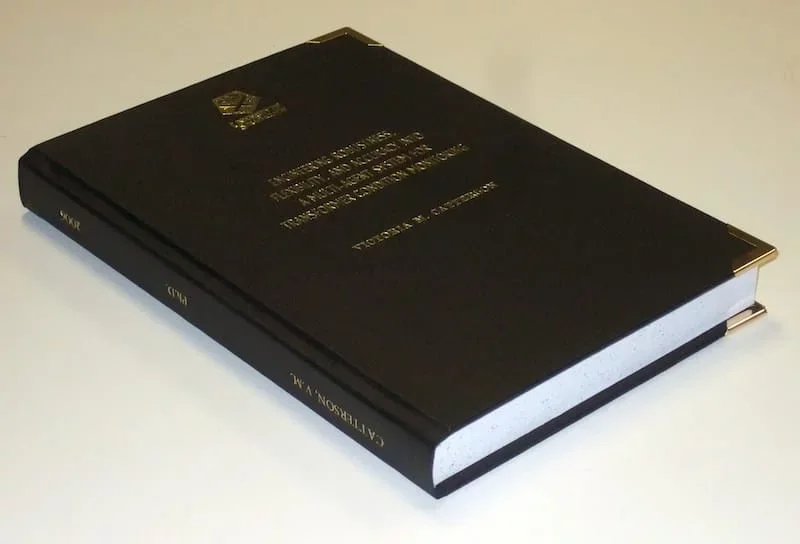
Image source: Thesis complete! (2018). Flickr. Retrieved 12 August 2018, from https://www.flickr.com/photos/cowlet/354911838 by Victoria Catterson
About The Author
Dr. amar udare, md, related posts ↓.
7 thoughts on “Radiology Thesis – More than 400 Research Topics (2022)!”
Amazing & The most helpful site for Radiology residents…
Thank you for your kind comments 🙂
Dr. I saw your Tips is very amazing and referable. But Dr. Can you help me with the thesis of Evaluation of Diagnostic accuracy of X-ray radiograph in knee joint lesion.
Wow! These are excellent stuff. You are indeed a teacher. God bless
Glad you liked these!
happy to see this
Glad I could help :).
Leave a Comment Cancel Reply
Your email address will not be published. Required fields are marked *
Get Radiology Updates to Your Inbox!
This site is for use by medical professionals. To continue, you must accept our use of cookies and the site's Terms of Use. Learn more Accept!
Wish to be a BETTER Radiologist? Join 14000 Radiology Colleagues !
Enter your email address below to access HIGH YIELD radiology content, updates, and resources.
No spam, only VALUE! Unsubscribe anytime with a single click.
- U.S. Department of Health & Human Services
- National Institutes of Health

En Español | Site Map | Staff Directory | Contact Us
- Science Education
- Science Topics
What is medical ultrasound?
How does it work, what is ultrasound used for, are there risks, what are examples of nibib-funded projects using ultrasound.
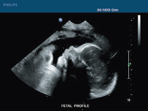
Medical ultrasound falls into two distinct categories: diagnostic and therapeutic.
Diagnostic ultrasound can be further sub-divided into anatomical and functional ultrasound. Anatomical ultrasound produces images of internal organs or other structures. Functional ultrasound combines information such as the movement and velocity of tissue or blood, softness or hardness of tissue, and other physical characteristics, with anatomical images to create “information maps.” These maps help doctors visualize changes/differences in function within a structure or organ.
Therapeutic ultrasound also uses sound waves above the range of human hearing but does not produce images. Its purpose is to interact with tissues in the body such that they are either modified or destroyed. Among the modifications possible are: moving or pushing tissue, heating tissue, dissolving blood clots, or delivering drugs to specific locations in the body. These destructive, or ablative, functions are made possible by use of very high-intensity beams that can destroy diseased or abnormal tissues such as tumors. The advantage of using ultrasound therapies is that, in most cases, they are non-invasive. No incisions or cuts need to be made to the skin, leaving no wounds or scars.
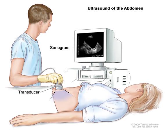
Ultrasound waves are produced by a transducer, which can both emit ultrasound waves, as well as detect the ultrasound echoes reflected back. In most cases, the active elements in ultrasound transducers are made of special ceramic crystal materials called piezoelectrics. These materials are able to produce sound waves when an electric field is applied to them, but can also work in reverse, producing an electric field when a sound wave hits them. When used in an ultrasound scanner, the transducer sends out a beam of sound waves into the body. The sound waves are reflected back to the transducer by boundaries between tissues in the path of the beam (e.g. the boundary between fluid and soft tissue or tissue and bone). When these echoes hit the transducer, they generate electrical signals that are sent to the ultrasound scanner. Using the speed of sound and the time of each echo’s return, the scanner calculates the distance from the transducer to the tissue boundary. These distances are then used to generate two-dimensional images of tissues and organs.
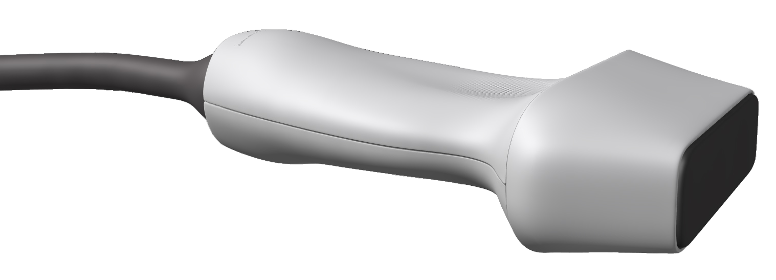
During an ultrasound exam, the technician will apply a gel to the skin. This keeps air pockets from forming between the transducer and the skin, which can block ultrasound waves from passing into the body.
Click here to watch a short video about how ultrasound works.
Diagnostic ultrasound. Diagnostic ultrasound is able to non-invasively image internal organs within the body. However, it is not good for imaging bones or any tissues that contain air, like the lungs. Under some conditions, ultrasound can image bones (such as in a fetus or in small babies) or the lungs and lining around the lungs, when they are filled or partially filled with fluid. One of the most common uses of ultrasound is during pregnancy, to monitor the growth and development of the fetus, but there are many other uses, including imaging the heart, blood vessels, eyes, thyroid, brain, breast, abdominal organs, skin, and muscles. Ultrasound images are displayed in either 2D, 3D, or 4D (which is 3D in motion).
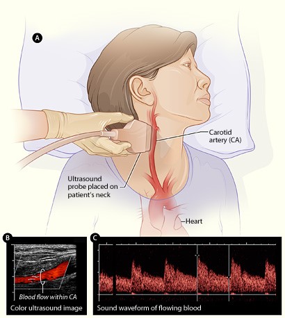
Functional ultrasound. Functional ultrasound applications include Doppler and color Doppler ultrasound for measuring and visualizing blood flow in vessels within the body or in the heart. It can also measure the speed of the blood flow and direction of movement. This is done using color-coded maps called color Doppler imaging. Doppler ultrasound is commonly used to determine whether plaque build-up inside the carotid arteries is blocking blood flow to the brain.
Another functional form of ultrasound is elastography, a method for measuring and displaying the relative stiffness of tissues, which can be used to differentiate tumors from healthy tissue. This information can be displayed as either color-coded maps of the relative stiffness; black-and white maps that display high-contrast images of tumors compared with anatomical images; or color-coded maps that are overlayed on the anatomical image. Elastography can be used to test for liver fibrosis, a condition in which excessive scar tissue builds up in the liver due to inflammation.
Ultrasound is also an important method for imaging interventions in the body. For example, ultrasound-guided needle biopsy helps physicians see the position of a needle while it is being guided to a selected target, such as a mass or a tumor in the breast. Also, ultrasound is used for real-time imaging of the location of the tip of a catheter as it is inserted in a blood vessel and guided along the length of the vessel. It can also be used for minimally invasive surgery to guide the surgeon with real-time images of the inside of the body.
Therapeutic or interventional ultrasound. Therapeutic ultrasound produces high levels of acoustic output that can be focused on specific targets for the purpose of heating, ablating, or breaking up tissue. One type of therapeutic ultrasound uses high-intensity beams of sound that are highly targeted, and is called High Intensity Focused Ultrasound (HIFU). HIFU is being investigated as a method for modifying or destroying diseased or abnormal tissues inside the body (e.g. tumors) without having to open or tear the skin or cause damage to the surrounding tissue. Either ultrasound or MRI is used to identify and target the tissue to be treated, guide and control the treatment in real time, and confirm the effectiveness of the treatment. HIFU is currently FDA approved for the treatment of uterine fibroids, to alleviate pain from bone metastases, and most recently for the ablation of prostate tissue. HIFU is also being investigated as a way to close wounds and stop bleeding, to break up clots in blood vessels, and to temporarily open the blood brain barrier so that medications can pass through.
Diagnostic ultrasound is generally regarded as safe and does not produce ionizing radiation like that produced by x-rays. Still, ultrasound is capable of producing some biological effects in the body under specific settings and conditions. For this reason, the FDA requires that diagnostic ultrasound devices operate within acceptable limits. The FDA, as well as many professional societies, discourage the casual use of ultrasound (e.g. for keepsake videos) and recommend that it be used only when there is a true medical need.
The following are examples of current research projects funded by NIBIB that are developing new applications of ultrasound that are already in use or that will be in use in the future:
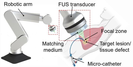
3D printing through the skin : Researchers at Duke University have developed a method to 3D print biocompatible structures through thick, multi-layered tissues. The approach entails using focused ultrasound to solidify a special ink that has been injected into the body to repair bone or repair soft tissues, for example. Initial experiments in animal tissue suggest the method could turn highly invasive surgical procedures into safer, less invasive ones. (Image on left courtesy of Junjie Yao (Duke University) and Yu Shrike Zhang (Harvard Medical School and Brigham and Women’s Hospital)).
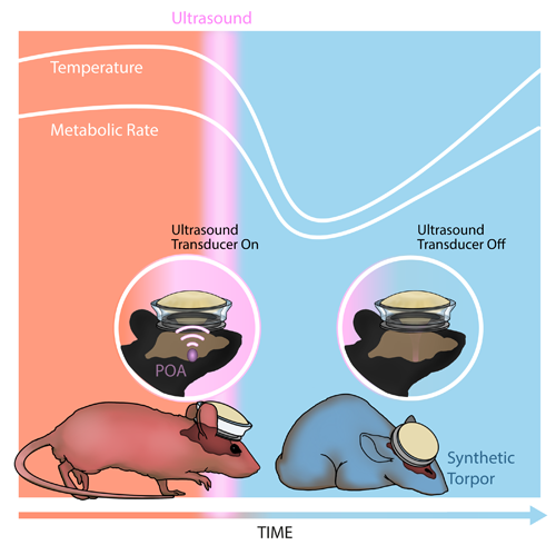
Inducing a hibernation-like state : Researchers at Washington University in St. Louis used ultrasound waves directed into the brain to lower the body temperature and metabolic rates of mice, inducing a hibernation-like state, called torpor. The researchers replicated some of these results in rats, which, like humans, don’t naturally enter torpor. Inducing torpor could help minimize damage from stroke or heart attack and buy precious time for patients in critical care. (Image on right courtesy of Yang et al./Washington University in St. Louis).
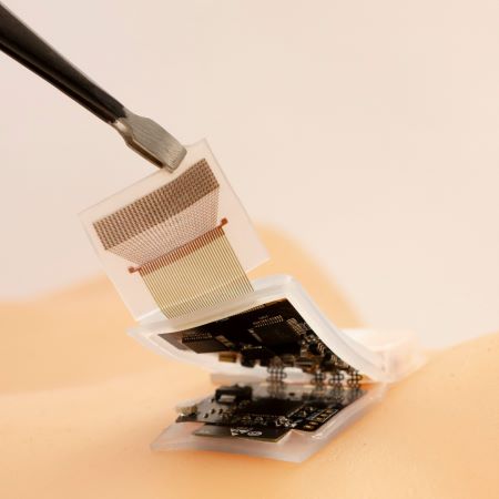
High-quality imaging at home : Brigham and Women’s Hospital researchers compared ultrasound scans acquired by experts with those taken by inexperienced volunteers, finding little difference in the image quality of the two groups. The unconventional approach of having patients take ultrasound images of themselves at home and share them with healthcare professionals could allow for remote monitoring and reduce the need for hospitalization. (Image on right courtesy of Duggan et al./Brigham and Women's Hospital).
Reviewed December 2023
Explore More
Heath Topics
- Breast Cancer
- Cardiovascular Disease
- Digestive Diseases
- Heart Disease
- Musculoskeletal Disease
- Reproductive Health
- Women's Health
Research Topics
- Optical Imaging
- Ultrasound (US) diagnostic
- Ultrasound (US) therapy
PROGRAM AREAS
- Ultrasound: Diagnostic and Interventional
Inside NIBIB
- Director's Corner
- Funding Policies
- NIBIB Fact Sheets
- Press Releases
Department of Circulation and Medical Imaging
- Master's programmes in English
- For exchange students
- PhD opportunities
- All programmes of study
- Language requirements
- Application process
- Academic calendar
- NTNU research
- Research excellence
- Strategic research areas
- Innovation resources
- Student in Trondheim
- Student in Gjøvik
- Student in Ålesund
- For researchers
- Life and housing
- Faculties and departments
- International researcher support
Språkvelger
Master thesis and projects - ultrasound technology - studies - department of circulation and medical imaging.
- Master thesis and projects
- Specialisation courses
Master's thesis and projects
Master's thesis and projects.
The Department of circulation and medical imaging offers projects and master's thesis topics for technology students of most of the different technical study programmes at NTNU. There is a seperate page for the supplementary specialisation courses .
List of topics
Topics for thesis and projects are given below. Most of the topics can be adjusted to the students qualifications and wishes.
Don't hesitate to take contact with the corresponding supervisor - we're looking forward to a discussion with you!
Asset Publisher
Blood flow imaging projects, estimation of true flow velocity using ultrasound, fusion of multi-modal cardiac data, pocket size ultrasound technology, pulse-echo based method for estimation of speed of sound, ultrasonic imaging through solids, surf imaging topics, ultrasound mediated drug delivery, real-time monitoring of left ventricular function under interventional procedures, fighting cancer with cw shear-wave elastography, adaptive clutter filtering for coronary heart disease, patient adaptive imaging in echocardiography, how to write ....
- a good abstract
- a good introduction
person-portlet
Lasse løvstakken professor.
Volume 5 Supplement 1
Topics in emergency abdominal ultrasonography
Edited by Luca Brunese and Antonio Pinto
Publication of this suppement has been funded by the University of Molise, Universiy of Siena, University of Cagliari, University of Ferrara and University of Turin. The Supplement Editors declare that they have no competing interests.
Sources of error in emergency ultrasonography
To evaluate the common sources of diagnostic errors in emergency ultrasonography.
- View Full Text
Accuracy of ultrasonography in the diagnosis of acute appendicitis in adult patients: review of the literature
Ultrasound is a widely used technique in the diagnosis of acute appendicitis; nevertheless, its utilization still remains controversial.

US detection of renal and ureteral calculi in patients with suspected renal colic
The purpose of this study was to determine whether the color Doppler twinkling sign could be considered as an additional diagnostic feature of small renal lithiasis (_5mm).
Gastrointestinal perforation: ultrasonographic diagnosis
Gastrointestinal tract perforations can occur for various causes such as peptic ulcer, inflammatory disease, blunt or penetrating trauma, iatrogenic factors, foreign body or a neoplasm that require an early re...
Sigmoid diverticulitis: US findings
Acute diverticulitis (AD) results from inflammation of a colonic diverticulum. It is the most common cause of acute left lower-quadrant pain in adults and represents a common reason for acute hospitalization, ...
The role of US examination in the management of acute abdomen
Acute abdomen is a medical emergency, in which there is sudden and severe pain in abdomen of recent onset with accompanying signs and symptoms that focus on an abdominal involvement. It can represent a wide sp...
Intestinal Ischemia: US-CT findings correlations
Intestinal ischemia is an abdominal emergency that accounts for approximately 2% of gastrointestinal illnesses. It represents a complex of diseases caused by impaired blood perfusion to the small and/or large ...
US in the assessment of acute scrotum
The acute scrotum is a medical emergency . The acute scrotum is defined as scrotal pain, swelling, and redness of acute onset. Scrotal abnormalities can be divided into three groups , which are extra-testicula...
Contrast enhanced ultrasound ( CEUS ) in blunt abdominal trauma
In the assessment of polytrauma patient, an accurate diagnostic study protocol with high sensitivity and specificity is necessary. Computed Tomography (CT) is the standard reference in the emergency for evalua...
Abdominal vascular emergencies: US and CT assessment
Acute vascular emergencies can arise from direct traumatic injury to the vessel or be spontaneous (non-traumatic).
Accuracy of ultrasonography in the diagnosis of acute calculous cholecystitis: review of the literature
To evaluate the accuracy of ultrasonography in the diagnosis of acute calculous cholecystitis in comparison with other imaging modalities.
Ultrasonography (US) in the assessment of pediatric non traumatic gastrointestinal emergencies
Non traumatic gastrointestinal emergencies in the children and neonatal patient is a dilemma for the radiologist in the emergencies room and they presenting characteristics ultrasound features on the longitudi...
- Editorial Board
- Sign up for article alerts and news from this journal
- Follow us on Twitter
- Follow us on Facebook
- ISSN: 2524-8987 (electronic)
Thank you for visiting nature.com. You are using a browser version with limited support for CSS. To obtain the best experience, we recommend you use a more up to date browser (or turn off compatibility mode in Internet Explorer). In the meantime, to ensure continued support, we are displaying the site without styles and JavaScript.
- View all journals
- My Account Login
- Explore content
- About the journal
- Publish with us
- Sign up for alerts
- Open access
- Published: 11 May 2024
A fully autonomous robotic ultrasound system for thyroid scanning
- Kang Su ORCID: orcid.org/0000-0002-3358-161X 1 na1 ,
- Jingwei Liu ORCID: orcid.org/0000-0002-3428-0210 1 na1 ,
- Xiaoqi Ren 2 , 3 na1 ,
- Yingxiang Huo ORCID: orcid.org/0000-0003-1197-5519 2 , 3 na1 ,
- Guanglong Du ORCID: orcid.org/0000-0001-9425-843X 1 na1 ,
- Wei Zhao 4 ,
- Xueqian Wang ORCID: orcid.org/0000-0003-3542-0593 5 ,
- Bin Liang 6 ,
- Di Li 7 &
- Peter Xiaoping Liu ORCID: orcid.org/0000-0002-8703-6967 8
Nature Communications volume 15 , Article number: 4004 ( 2024 ) Cite this article
Metrics details
- Computer science
- Echocardiography
- Thyroid diseases
The current thyroid ultrasound relies heavily on the experience and skills of the sonographer and the expertise of the radiologist, and the process is physically and cognitively exhausting. In this paper, we report a fully autonomous robotic ultrasound system, which is able to scan thyroid regions without human assistance and identify malignant nod- ules. In this system, human skeleton point recognition, reinforcement learning, and force feedback are used to deal with the difficulties in locating thyroid targets. The orientation of the ultrasound probe is adjusted dynamically via Bayesian optimization. Experimental results on human participants demonstrated that this system can perform high-quality ultrasound scans, close to manual scans obtained by clinicians. Additionally, it has the potential to detect thyroid nodules and provide data on nodule characteristics for American College of Radiology Thyroid Imaging Reporting and Data System (ACR TI-RADS) calculation.
An AI-powered navigation framework to achieve an automated acquisition of cardiac ultrasound images
Radiomics feature reproducibility under inter-rater variability in segmentations of CT images
Face mediated human–robot interaction for remote medical examination
Introduction.
Ultrasound (US) diagnosis is widely used in examining organs, such as the liver, gallbladder, pancreas, spleen, kidney, and thyroid 1 , 2 , 3 , 4 , 5 , 6 . However, the qualities of US diagnosis rely heavily on the experience and skills of the sonographer and radiologist 7 , 8 , 9 . The acquisition of US images usually exhibits variability among clinicians, and even the same examiner may potentially produce very different results from different scans 10 . Furthermore, the standard practice of having patients to assume a supine position and maintain still- ness during examinations can present challenges, as these requirements may not always be met 11 . Consequently, operator-dependency and patient-specific factors introduce inconsistency and unreliability into US diagnosis results 12 .
From pure human control to complete autonomy, the level of autonomy of medical robots can be classified into different categories 13 , 14 . According to the framework presented in ref. 13 , the level of autonomy includes level 0, which is defined as no autonomy, e.g., tele-operated systems or prosthetic devices; level 1, robotic assistance, the robot guides the human during a task while the human maintains a continues control; level 2, task autonomy, the robot provides discrete rather than continuous control over a specific task; level 3, conditional autonomy, the system is capable of generating different task strategies but relies on the human’s selection or approvement; level 4, high autonomy, the system is capable of making medical decisions but only when supervised by a qualified physician; level 5, full autonomy, the robot can perform the entire procedure without any human involvement. Under the concept of the autonomy level of medical robots (Level 0–5), the autonomy level of ultrasonic inspection robots (Level 0–3) has been defined at present. Level 0 is defined as “manual probe manipulation”, the proposed tele-echography systems 15 , 16 , 17 , 18 , 19 . The US robotic system in Level 1 utilizes visual servo technology to allow the robot to automatically track desired image features 20 , 21 , 22 , 23 and compensate for unnecessary patient movement during remote operation. Level 2 is described as performing autonomous US acquisition along a manually planned path 24 , 25 . The US robotic system in Level 3 can autonomously plan and perform US acquisition without any instruction from a human operator but requires the supervision of an operator 26 , 27 , 28 , 29 .
With the improvement of the autonomy of ultrasonic inspection of robots, substantial advancements in the field have been achieved, providing promising solutions to improve the accuracy and efficiency of US procedures. The prerequisite for robotic US acquisitions is to plan the scanning path to ensure finding a desired imaging plane or covering a selected region of interest. In general, existing systems typically rely on global information of the target tissue acquired from preoperative medical images or surface information obtained from external sensors 27 , 30 , 31 . Given the inherent variability in individual anatomy and the dynamic of human motion, executing a scanning task based on a predetermined trajectory presents significant challenges. To address these difficulties, Jiang et al. 32 integrated the feedback of segmented images into the control process. Zhan et al. 33 have proposed a visual servoing framework for motion compensation. However, these methods usually assume that the target features exist in the US image, and once the features are lost, the control methods may fail. In an effort to apply online image-guided methods to define the scanning trajectory and derive clinically relevant information out of the 3D reconstructed image, Zielke et al. 34 implemented in-plane navigation specifically designed for robotic sonography in thyroid volumetry. However, in actual clinical practice, sonographers often use a combination of multiple views, such as transverse and longitudinal scans, for the identification and diagnosis of both benign and malignant pathologies 35 . Furthermore, the increased autonomy of the US robotic system may lead to a higher risk of injury to patients due to machine failure, so the clinical effectiveness of the system needs to be further studied. Although many implementations have been proposed 36 , 37 , 38 , 39 , 40 , the overall success scanned rate is low due to the differences between human bodies. To this end, the fully autonomous robotic diagnosis system adapted to clinical practice is still challenging, as it calls for more perception, planning, and control on the part of the robot while taking into account patient safety.
In order to eliminate the above obstacles, we developed a fully autonomous robotic ultrasound system (FARUS), as shown in Fig. 1 . To the best of our knowledge, this is the first in-human study of fully autonomous robotic US scanning for thyroid. In conventional US examinations, the process involves a division of responsibilities between sonographer and radiologist. However, the presented FARUS integrates the both roles into a single autonomous unit. Here, we achieved a human-like fusion of both in-plane and out-of-plane scanning, allowing for comprehensive scanning of the thyroid region, and providing a detailed evaluation of the anatomy. The FARUS overcomes the challenges associated with the localization of thyroid targets through a reinforcement learning strategy. It enables to optimize the orientation of the probe based on Bayesian optimization. It also uses deep learning techniques for real-time segmentation of the thyroid gland and potential nodules. As a result, this system provides a convenient autonomous tool integrating nodule detection, lesion localization and automatic classification.
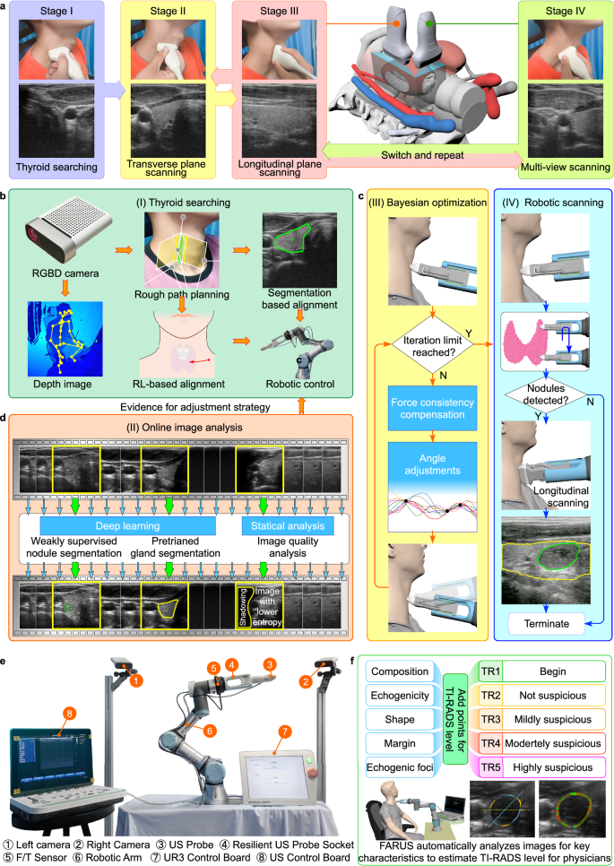
a The four-stage thyroid scan procedure used by clinical doctors. Stage I: the doctor placed a US probe below the thyroid cartilage and found the thyroid lobe in the US image; Stage II: the doctor performed IPS from the breastbone to the hyoid bone, and backward; Stage III: the doctor performed OPS to screen for thyroid disease; Stage IV: the doctor checked multi-view of the thyroid. b – d Control architecture of the full autonomous control strategy for robotic thyroid ultrasound imaging. Initially, we plan the preliminary scanning path through the human skeleton and subsequently complete the thyroid search process using reinforcement learning and thyroid segmentation. Gland and nodule identification are performed using a pretrained gland segmentation model and a weakly supervised nodule segmentation model, respectively. Throughout the scanning process, we used Bayesian optimization to adjust the scanning angle. Additionally, we combined IPS and OPS to perform multi-angle scanning for suspected nodule areas. e The overview of the experimental setup. f FARUS enables estimate TI-RADS level with key characteristics of nodules.
The second contribution of this work lies in the practical realization and clinical application of the FARUS, which achieves high-quality US images in comparison with those manually collected by sonographers, and realizes accurate and real-time detection of thyroid nodules. We investigated the validity of our approach by conducting a comparative evaluation of FARUS-driven diagnostic results for thyroid nodules against the established hospital benchmark. We have conducted extensive evaluations and studied the system’s performance and reliability. Our work addresses the gap between existing research and clinical application by demonstrating the deployment of this system in a real-world clinical setting.
System design for autonomous ultrasound imaging
The robotic scanning procedure comprises four phases, mirroring the clinical workflow: thyroid searching (TS), in-plane scanning (IPS), out-of-plane scanning (OPS), and multi-view scanning (MVS), as depicted in Fig. 1a . An overview of our autonomous robotic system for thyroid scanning and real-time analysis is presented in Fig. 1e . The system consists of a six-degree-of-freedom UR3 manipulator that carries a linear US probe, a US probe fixture and a six-axis force/torque sensor. The high-frequency 2D linear US probe enables the optimal depth penetration within the superficial location of the thyroid tissue. The six-axis force/torque sensor can detect three orthogonal forces and torques between the human neck and probe. The Kinect camera tracks 3D view skeleton joints of the human body, while its 2D view provides visual feedback for the operator supervising the robotic system. It is remarkable that the entire scanning process, including thyroid searching, force control, image quality optimization, and suspected nodule detection was completed autonomously.
The following describes the thyroid scanning workflow of the proposed FARUS, as in Fig. 1b–d . First, the participant was instructed to turn his/her head after applying US gel to the neck skin. The scanning range was specified as a rectangular area with a length of 6.47 cm and a width of 5.48 cm. The contact force within the range of 2.0 N and 4.0 N was maintained to ensure sufficient pressure and prevent pressure-induced shape distortion of the thyroid anatomy. The thyroid search procedure began when the probe reached the estimated position given by skeleton joint locations. Due to individual anatomical variations, the thyroid gland may not be immediately visible in the US image obtained at the probe’s estimated position. In such case, we used reinforcement learning to adjust the probe’s movement until the thyroid gland is accurately located. The search procedure was finished when the gland region, segmented by our gland segmentation model, exceeds a predetermined threshold. Subsequently, the probe orientation was optimized through Bayesian optimization 41 , see Fig. 1c . In the IPS procedure, the probe scans upward until the upper thyroid lobe end is invisible, then scans downward. Nodule locations are recorded during detection by our segmentation network. Out-of-plane scanning is employed for previously recorded nodules while avoiding clavicle and jaw collisions. The scanning halts if FARUS detects significant participant movement. Finally, we use the ACR TI-RADS scoring method to classify nodules as either benign or malignant, based on their distinct characteristics, see Fig. 1f .
Deep learning for gland and nodule segmentation
For a fully autonomous robotic thyroid scan, real-time location information of thyroid lobes and nodules is crucial. Given the stable characteristics of healthy thyroid lobes and the diverse nature of nodules, we used two separate networks for the thyroid lobe and nodule segmentation tasks, respectively, as in Fig. 2a . Each model incorporates a pre-trained encoder for feature extraction and employs the UNet 42 architecture for the decoder, generating masks from extracted features. To train the nodule segmentation network with prior knowledge from the thyroid lobe mask, thyroid lobe pseudo-labels are generated for nodule images. Spatial and feature constraints are then applied to enhance nodule segmentation based on these pseudo-labels. The spatial constraint ensures proximity to the thyroid lobe, while the feature constraint emphasizes regions with similar gray values. To address overfitting with limited samples, a two-step approach is employed: pre-training on a nodule source dataset followed by fine-tuning on our smaller dataset.
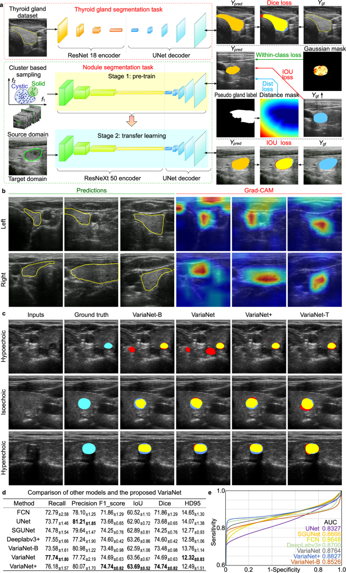
a Thyroid and nodule segmentation network based on pre-training and weakly supervision. The feature and spatial losses are used to provide prior knowledge to the network considering the diversity of nodule samples and the space constrain between thyroid lobes. b Thyroid lobe segmentation based on pre-training. c Our proposed VariaNet and its variants predict results for different types of nodules. d Comparisons with the existing segmentation models on the SCUTN1K testset (best result in bold). e Receiver Operating Characteristic (ROC) curve of different algorithms on the SCUTN1K testset. Source data are provided as a Source Data file.
To understand the segmentation preferences of the gland segmentation model across different slices, we used Grad-CAM 43 to visualize the fourth level output of the encoder. In Fig. 2b , the highlighted regions are more closely aligned with the areas of interest during central thyroid scanning. However, when scanning the upper pole of the thyroid, the attention map reveals other targets, such as muscles. To address cases where the thyroid target is small or even invisible, we introduced reinforcement learning to the thyroid search process, see “Thyroid search and probe orientation optimization” section. Figure 2c shows the segmentation results of our proposed VariaNet on three types of thyroid nodules. Here, VariaNet-B, VariaNet, and VariaNet+ denote models trained with different types of loss functions: no additional loss, feature loss only, and a combination of feature and spatial loss, respectively. VariaNet-T represents the refined model, trained with a dual loss function and source domain data. The integration of distance loss was critical to constrain hypoechoic nodules, which often have similar gray value distributions to other tissues, and thereby minimize the false positive rate. At the same time, the implementation of feature loss improved the segmentation performance on isoechoic nodules, especially in cases where the gray values of the nodules closely match those observed in the thyroid region. In addition, the application of transfer learning augmented with prior knowledge proved to be effective in strengthening the robustness of the network, as evidenced by the segmentation of hyperechoic nodules. In Fig. 2d , a comparative evaluation between our proposed VariaNet and other semantic segmentation models shows that the fusion of weakly supervised learning improves the segmentation capabilities of the nodule network. The proposed VariaNet exceeds the baseline model VariaNet-B by 0.97% IoU score on the SCUTN1k testset. We present ROC curves to intuitively illustrate the performance of the proposed method, proving that VariaNet outperforms other existing methods due to the tailor-made loss function, see Fig. 2e .
Thyroid search and probe orientation optimization
An important problem in robotic thyroid scanning is the localization of the thyroid on the body surface. We present a coarse-to-fine approach to thyroid localization. In the coarse estimation step, the neck region is identified by human skeletal key points. Notably, the thyroid lobe may not be visible based on the location predicted by the skeletal data, because the anatomy of the neck varies widely in different populations. Therefore, we added the fine-tuning step to further localize the thyroid lobe. To enable robotic scanning in such a case, we used reinforcement learning to determine the location of the thyroid.
Figure 3 shows the training process of Deep Q-Network (DQN) learning with panoramic environment. The process starts with sequences of data that are collected and labeled manually. We labeled each sequence of thyroid images a goal or an ideal position for model to learn as shown in Fig. 3a . After that, each of sequence of image will be aligned and attached to be a panorama as shown in Fig. 3b . Then a bunch sequence of image will be randomized and generated into panoramas as shown in Fig. 3c, d . There is a sliding window will slide according to the action given by the agent. To mimic the real environment when sometimes the probe is not fully attached, we combine simulated view with the random noise as shadow mask, to generate imperfect thyroid images that mimic the appearance when the probe is not fully attached, as shown in Fig. 3e . Figure 3f presents the results of the training evaluation for reinforcement learning. During the exploration stage, which comprises the initial 30,000 steps. Figure 3g illustrates a progressive increase in the mean reward, indicating the gradual improvement of the RL model’s performance on the given task as it continues to learn over time. Figure 3h depicts the process of thyroid scanning facilitated by the DQN Learning model in the context of FARUS. The DQN learning model enables the robotic arm to execute movements based on the input received from US images. As illustrated in Fig. 3h , the model accurately predicts the required movements to guide the robotic arm effectively.
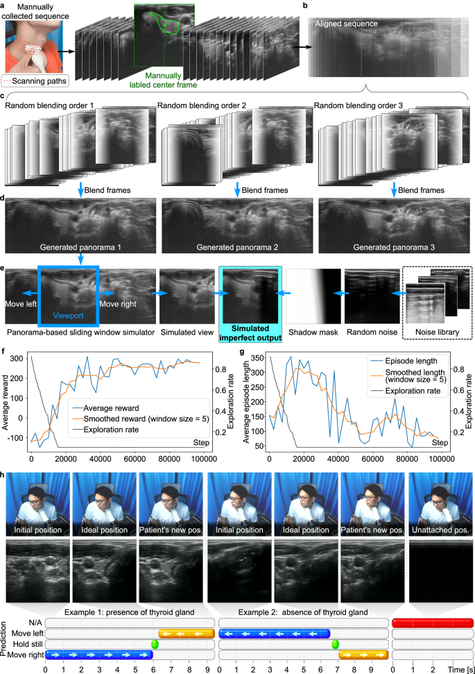
a A sequence of image is collected and labeled manually. b The alignment of sequence image into panorama. c Blending images in a random sequence. d Generated panorama. e The simulation of panorama-base sliding window. f Average reward vs. step. g Average episode length vs. step. h Trained model’s prediction vs. time (please refer to Supplementary Movie 1 for additional details). Source data are provided as a Source Data file.
In the first and fourth images, the probe is first attached to the patient’s neck. The model then predicts the appropriate directions for the robotic arm to move in each frame. The blue bar represents the model’s predictions for movement to reach the ideal position. The green bar represents the model’s prediction for maintaining a stationary position once the ideal position is reached. Conversely, the yellow bar represents the model’s predictions when the patient is already in motion. The DQN learning model instructs the robotic arm to move to the right based on the first thyroid image until the arm reaches the position with the ideal thyroid image, as shown in the second thyroid image. Notably, our proposed FARUS can effectively guide the robotic arm even when the patient is moving. For example, at 6.3 s, when the patient’s neck moves to the left, the model adjusts the position of the robotic arm accordingly, resulting in the leftward movement shown in the third thyroid image. In the fourth thyroid image, the robotic arm is attached to the mid-neck region, which is not the correct location to detect the presence of the thyroid gland in the neck. The model guides the robotic arm to move to the left until it reaches the ideal position. In addition, even in the absence of thyroid presence in the fifth image, the model has learned to predict the ideal position and guides the robotic arm to stop at that position. The DQN learning model in FARUS demonstrates its ability to accurately guide the robotic arm during thyroid scanning, even in the presence of patient movement or the absence of the thyroid gland.
Experienced sonographers usually fine-tune the probe angle after locating the thyroid to obtain high-quality US images. To imitate such an expert behavior, an autonomous robotic system should assess the quality of US images and feed them back to adjust the probe orientation. However, taking into account the limited resolution of the Kinect and potential participant movements during the scan, the pre-estimated normal vector cannot be directly applied as the normal vector for subsequent scans. To tackle this problem, Bayesian optimization algorithm with image entropy as the loss function was used to obtain a better probe orientation with very few adjustments. Although some statistical methods such as grayscale, confidence map and root mean square error (RMSE) have been proposed, there is still no gold standard for evaluating US image quality 44 . In this work, we used the image entropy to evaluate US image quality because it is highly effective for image processing and can be used to assess texture in images based on a statistical measure of randomness 45 .
In many instances, Bayesian optimization (BO) outperforms expert as well as other state-of-the-art global optimization algorithms. Bayesian optimization constructs a surrogate model for the objective function and quantifies the uncertainty through Bayesian inference. This surrogate model determines where the next candidate will be, as in Fig. 4a . The significant differences in image entropy values were observed after the BO phase for 89 participants (Fig. 4b ). During the BO phase, the position of the US probe remains constant; therefore, probe orientation and contact force are the two major factors in the entropy value of the US image. We consider a limited budget of N = 5 iterations to speed up the BO phase. The entropy of the US image varies from 7.172 to 7.173 in 5 iterations, and the max entropy corresponding to the optimal orientation was reached at the second iteration (Fig. 4c, d ). When the number of iterations reached 5, a higher drop in performance was observed due to its exploration nature. During this experiment, the probe was initially set at a relatively optimal angle, resulting in a less noticeable entropy increase before and after Bayesian optimization. However, between 21.3 and 24.8 s, the US probe underwent a 10-degree angle adjustment, revealing that the entropy value of the US image responded more sensitively to angle changes compared to image confidence. This sensitivity could be attributed to the level of contact between the skin and the probe. As illustrated in Fig. 4e , there is a positive relationship between the image entropy and confidence map 46 that evaluates the contact condition at each pixel. As the entropy value consumes less computation, it allows for a real-time control of image quality. To further explore the influence of contact force on image quality, we conducted an investigation involving nine participants. Each participant was instructed to apply varying levels of force on the US probe, positioned at the robot’s end, while ensuring safety under continuous manual monitoring. Figure 4f demonstrates that when the contact force between the probe and the skin exceeds 2 N, the median of the image entropy value remains stable. Taking scanning comfort and safety into account, we set the maximum contact force at 4 N.
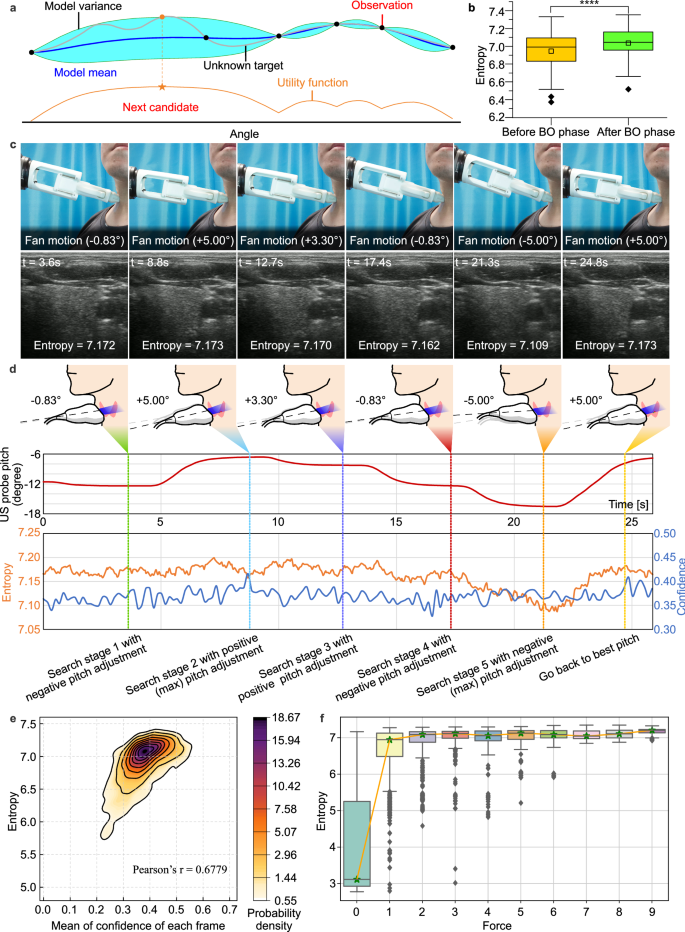
a Overview of Bayesian optimization. Example illustrating a Gaussian process surrogate model fitting to data derive from a unknown target and its expected utility function maximizing to select the next candidate. b The boxplot displaying the significant differences in image entropy values after BO phase for participants ( n = 89); the top, middle, and bottom boundaries of the boxplots represent 25th, 50th, and 75th percentile, respectively; the small squares represent the mean. Outliers defined when value larger than 1.5*IQR + 75th percentile; **** p < 0.0005, p value obtained with a paired, two-sided t -test. c , d Probe orientation optimization procedure shown the five decisions made by BO with image entropy as loss function (please refer to Supplementary Movie 2 for additional details). e The relationship between image entropy and mean confidence 46 that characterize the contact condition of US image, as seen in a positive correlation between two evaluation metrics. f Force values greater than 2 N do not cause major changes in the median entropy. The 13,256 pairs of data from 9 participants ( n = 9) were divided into 10 groups and playback sampled 800 times for each group; the top, middle, and bottom boundaries of the boxplots represent 25th, 50th, and 75th percentile, respectively; the stars represent the median. Outliers defined when value larger than 1.5*IQR + 75th percentile. Source data are provided as a Source Data file.
Fully autonomous robotic ultrasound imaging
In actual clinical practice, sonographers frequently employ a combination of multi-view scanning methods to conduct comprehensive and detailed diagnosis of suspected nodules. Drawing inspiration from this clinical experience, once FARUS finds a suspected nodule in the transverse scan, the longitudinal scan will also be performed for further investigation. As displayed in Fig. 5a , we have achieved full autonomous scanning based on the fusion of force and visual information. Figure 5b shows the robotic scanning procedure for the right thyroid lobe of one participant. During the transverse scanning, we can see how the thyroid area increases first and then decreases, beginning with the appearance of the upper pole and ending with the disappearance of the lower pole. Moreover, shadows were avoided and thyroid lobes were centered with control strategy. The contact force, probe position and probe orientation are shown in Fig. 5c . As illustrated in Fig. 5c , noticeable fluctuations in the force value are observed during the initial 20 s of the transverse scan. This phenomenon arises as the US probe transitions from the middle of the thyroid toward the upper pole, following Bayesian optimization. The contact between the probe and the skin is affected by the surrounding thyroid cartilage at the upper pole, causing an unstable change in force during this movement. A similar instability is also observed during the scanning of the lower pole of the thyroid. Analysis of the probe’s position change reveals that FARUS maintains a constant speed in the Z -axis direction throughout the transverse scanning process. However, non-uniform adjustments occur in the Y and X directions, which are related to centering the thyroid gland. In the longitudinal scanning process, if multiple nodules are detected, their image features and location information are combined to determine whether these nodules were scanned during the transverse scanning process. This determination is achieved by matching the position of the nodules within the thyroid.
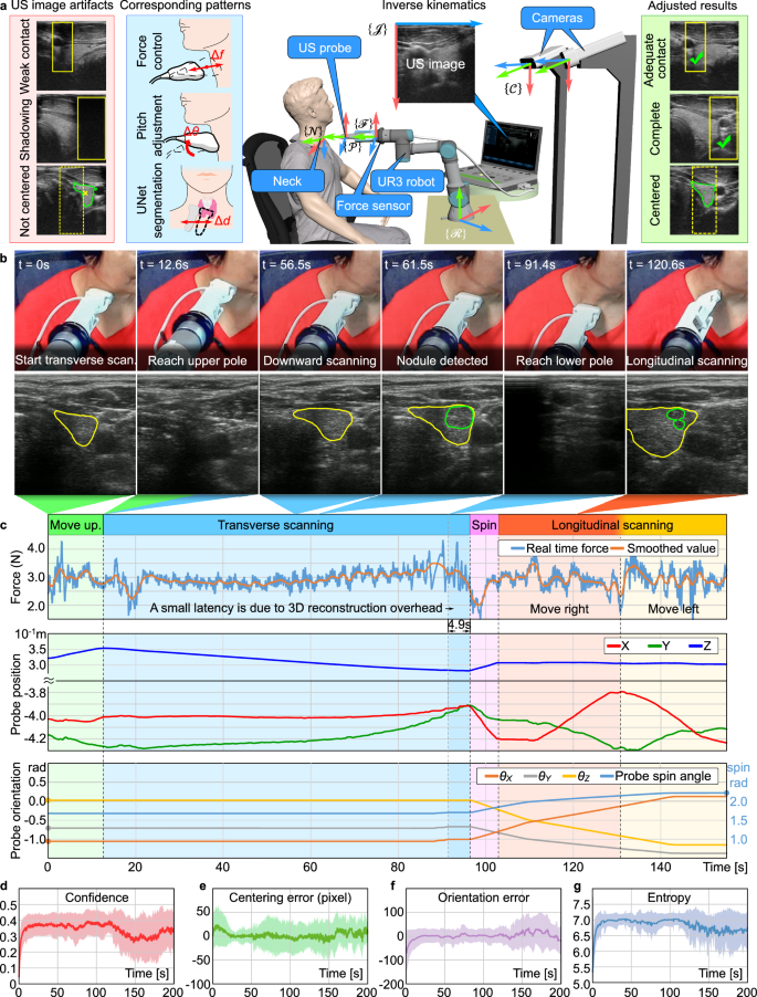
a The control strategy of FARUS. The autonomous scanning is achieved based on the fusion of force and visual information. b , c Evolution of force, probe position, and probe orientation for a patient with nodules during the experiment, which included the transverse scanning phase and longitudinal scanning phase. During the transverse scanning phase, the orientation of the probe at the end of the robot remained unchanged. Guided by the thyroid gland segmentation network, FARUS maintained the thyroid lobe in the center while moving the probe from the upper pole to the lower pole of the thyroid. When the nodule segmentation network detected a suspected nodule, the location of the suspected nodule was recorded. In the longitudinal phase, FARUS re-scanned the suspected nodules from a different angle to determine whether they were lesions (please refer to Supplementary Movie 3 for additional details). d – g Performance of the scanned image quality of FARUS on 19 patients. The performance metrics for evaluating the US image quality included confidence map, centering error, orientation error and image entropy. The shadowed area represents mean ± SD over the different experiments, while the curves inside the shadowed areas are the average results. Source data are provided as a Source Data file.
For the evaluation of the US image quality, four evaluation metrics including confidence map 46 , centering error, orientation error and image entropy were used to characterize the contact condition, thyroid visibility, orientation performance, and texture details of US image, respectively. From Fig. 5d–g , in the first 20 s, the entropy value and confidence value of the image increase as the probe makes contact with the patient, while the centering error and orientation error decrease. After 25 s, the mean value of the centering error of the image decreases to 0, indicating the completion of the thyroid search process and the image centering process. In the subsequent Bayesian optimization process, the entropy increase point is not distinctly visible due to variations in the optimization time for each patient. The centering error remain relatively stable between 25 and 100 s. After 150 s, the mean square error of confidence map, orientation error and image entropy increases, which is associated with the additional OPS phase.
ACR TI-RADS risk stratification using FARUS
In this study, we present the FARUS system, designed for scanning, detection, and classification of nodules in a sample of 19 patients. The ACR TI-RADS 47 is employed for nodule classification based on their US characteristics. Additionally, we conduct a comparative analysis between FARUS-generated classifications and evaluations provided by professional sonographer. Figure 6a displays five nodules that demonstrate complete agreement with the sonographer’s diagnosis. Results from the scoring and classification process by FARUS, following ACR TI-RADS criteria, are in Fig. 6b . To assess the echogenicity, composition, and echogenic foci of the thyroid nodule, we analyze the distribution of pixels in the thyroid gland and the nodule (Fig. 6c ). In Fig. 6b , the nodule’s composition is categorized as solid, leading to a score of 2, while its echogenicity is classified as hypo, also resulting in a score of 2. The nodule demonstrates a well-defined boundary and a regular shape, contributing to a score of 0. Additionally, the comparison of height and weight exceeds 1, resulting in a score of 3. Lastly, no echogenic foci are observed within the nodule, leading to a score of 0. The total score amounts to 7, leading to the classification of the nodule as level 5 or highly suspicious. Another example of a nodule has a mixed composition, leading to a score of 1, and its echogenicity as hypo, resulting in a score of 2. The nodule’s characteristic features, including a clear boundary and regular shape, warrant a score of 0 points. Additionally, the comparison of height and weight yields a score of 0 as it is less than 1. Furthermore, no presence of peripheral calcification within the nodule leading to a score of 0. Consequently, the cumulative score amounts to 3, classifying the nodule as level 3 or mildly suspicious.
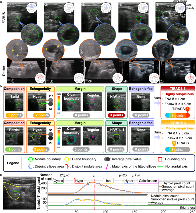
a Indicative nodules identified with FARUS and the respective US images provided by doctors. b Examples of two separate nodules, with accompanying explanations of their TIRADS scores. c Correlation between number of pixels over brightness in thyroid image. Source data are provided as a Source Data file.
According to doctor’s evaluation, 17 individuals were found to have nodules, while 2 individuals showed no presence of nodules. Our developed system, FARUS, identified 13 individuals as having nodules and 6 individuals as having no nodules. The scoring and recommended management of 24 nodules among the 13 individuals detected by both FARUS and the doctor were compared. Each nodule detected by FARUS was matched to the doctor’s report based on its location and shape. Table 1 shows the comparison between FARUS and doctor scoring and the classification of thyroid nodules (please refer to Supplementary Data 1 for more details). Among the 24 nodules assessed, 10 were diagnosed with the same score distribution by both the doctor and the developed FARUS. This can be attributed to the US image’s appropriate brightness and contrast, resulting in well-distributed pixels. Additionally, 8 nodules exhibited a score difference of 1, primarily attributed to discrepancies in echogenicity or composition classifications. There were four nodules with a score difference of 2 and another with a score difference of 4, possibly caused by different classifications in both echogenicity and composition. Nodule size variations in the scan may be attributed to differences in patient positioning: the doctor scans in a supine position, while FARUS scans in an upright sitting position due to security concerns. The doctor’s report indicates that out of the 19 patients, one patient has two nodules requiring fine-needle aspiration (FNA) and follow-up, respectively. For this patient, FARUS results were consistent with the doctor’s assessment. In the cases of the nodule #5, diagnostic inconsistency arose from variations in echo characteristics, leading to divergent recommended management.
The main reason for the different classifications by FARUS is the use of a probe different from the one used by the doctor. The doctor’s probe consistently produces a specific color of the thyroid gland for each patient, while the brightness and contrast of US images produced by our probe may vary across different patients with varying ages and weights. The proposed FARUS system classified nodules, with complete agreement in 10 out of 24 cases with the doctor’s diagnosis. Furthermore, for the remaining nodules, the discrepancies were primarily limited to 1 score difference, mainly arising from variations in echogenicity and composition. Current volunteer participants typically have low-risk nodules and clinical FNA is not recommended. Although FARUS has demonstrated feasibility and potentiality in nodule detection and data collection for ACR TI-RADS classification, further clinical studies are essential to assess its safety as a screening tool for probably or definitely malignant nodules.
Current US examination relies on sonographers to perform scanning operations. Patients often need to make an appointment for US examinations, resulting in long waiting times and often delays in treatment. At the same time, last decades have seen a rise in thyroid nodules. The detection and diagnosis of thyroid nodules still rely heavily on the expertise and experience of doctors. Compared with the traditional manual diagnosis or remote diagnosis, this fully autonomous robotic system adopts a patient-centered concept, allowing the patient to be examined in a more comfortable way. Furthermore, FARUS is suitable for rapid screening in out-patient clinics and remote low-level centers as it enables scan autonomously without intervention.
In this study, we developed an automated US diagnostic robot with artificial intelligence, which can accurately diagnose thyroid nodules. It is expected to be equipped not only in specialized hospitals, but also in clinics and remote areas. This non-invasive, rapid, and accurate screening strategy can provide an early warning of thyroid nodule development. The system operates on an autonomous scanning mode, a notable advantage of which is the no need of direct contact between medical staff and patients. This configuration effectively minimizes the risk of transmitting infectious diseases between patients and healthcare providers.
Currently, most US robotic systems studies do not include comparisons with doctors, nor do they evaluate participants’ satisfaction. As shown in Table S1 , we recruited 14 sonographers from 7 hospitals to make evaluations of our acquired transverse and longitudinal images, assigning scores quantified into five levels ranging from 1 to 5. Specifically, a score of 1 denoted “very poor”, 2 denoted “poor”, 3 denoted “medium”, 4 denoted “good”, and 5 denoted “very good” image quality. It shows that the images acquired by the FARUS have good quality, centrality and integrity. According to Fig. 7 , most participants who took part in the scanning felt safe with our system and experienced no adverse reactions, such as pain and other discomfort. Certain participants expressed feelings of anxiety regarding the robot scan and harbored concerns regarding its safety. The majority of the participants expressed that US robots cannot replace doctors, primarily due to doctors’ possession of a vast repository of medical knowledge, which the robots lack at the current moment. Moreover, doctors occupy a pivotal position as esteemed medical experts and trustworthy sources of aid.
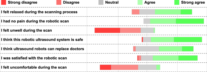
A clear trend of agreement or disagreement is more obvious when the entire bar (100%) is shifted left or right ( n = 70). Source data are provided as a Source Data file.
To compare the image quality of robotic US scans with that of manual scans, we invited five doctors with between 3 and 15 years of experience to scan the thyroids of 13 participants. In Fig. 8a , the FARUS completed transverse and longitudinal scanning in the same manner as a doctor. In the OPS phase, FARUS can detect suspected nodules as well as cover the carotid arteries. During the IPS phase, the FARUS was able to scan a continuous area from the upper pole to the lower pole of the thyroid gland, while ensuring that the thyroid gland was centered. As shown in Fig. 8b–e , there is still variation among doctors when using entropy, center error, mean confidence, and left-right intensity symmetry (LRIS) as image evaluation metrics. Specifically, LRIS refers to the ratio of gray scale distribution between the left and right sides of US images. Therefore, we performed a comparative analysis of five doctors as a group with robot performance. Figure 8f–i shows some similarity in the evaluation metrics between the robot and the doctors for both the IPS and OPS phase. The centering error of the IPS phase of robotic US scans is smaller than that observed in scans conducted by five doctors. This difference can be attributed to the robot control process. Additionally, differences in scanning method and range may also contribute to differences in entropy value, mean confidence, and image grayscale distribution between robot and doctors.
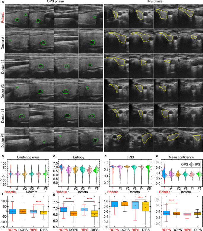
a The in-plane/out-of-plane US image sequences acquired by the FARUS and five experienced doctors on the same two participants. The green contour in the US images represent the segmented suspicious nodules by our proposed VariaNet, while the yellow contour represent the segmented thyroid lobe by our pre-trained gland segmentation model. b – e Violin plots illustrating the four evaluation metrics between FARUS and five doctors. Dashed line indicates median; dotted lines indicate 25th and 75th percentiles; n = 13 participants. f – i Boxplots displaying the four evaluation metrics between FARUS and five doctors. ROPS robotic out of-plane scanning, DOPS doctor out-of-plane scanning, RIPS robotic in-plane scanning, DIPS doctor in-plane scanning, LRIS left–right intensity symmetry. The top, middle, and bottom boundaries of the boxplots represent 25th, 50th, and 75th percentile, respectively; the small squares represent the mean. Outliers defined when value larger than 1.5*IQR + 75th percentile; n = 13 participants; p value obtained with bootstrap tests; **** p < 0.0005. Source data are provided as a Source Data file.
We also compared the probe motion of the robotic scanning with that of manual scanning. The probe motion data during doctors’ scanning process were recorded with a probe motion sensor. As compared to manual scanning, the robotic scanning was more stable in terms of force and velocity, as in Table S2 . In general, humans were more efficient than FARUS. The FARUS took 213.0 ± 85.3 s to complete a single thyroid lobe scan for 70 participants, while five doctors spent 67.2 ± 27.6 s to complete the scan for 13 participants. These differences can be attributed to dynamic path planning and force-controlled feedback, the former to compensate the motion and the latter to ensure participants’ comfort. Moreover, a conservative speed is implemented in our FARUS system to ensure the safety of participants.
The FARUS comprises three primary stages: scanning, detection, and classification, each of which plays a crucial role in influencing FARUS’s performance. During the detection stage, the main challenge faced by FARUS in detecting nodules is primarily attributed to the size and echogenicity of the nodules. Table 2 shows missed and possible false positive thyroid nodules detected by FARUS. The results of this study indicate that smaller nodules, such as #25, #26, #27, #28, #29 and #30 present greater difficulty for FARUS to detect. Similarly, nodules with isogenic properties, such as #31 and #32, also pose challenges for FARUS’s detection capabilities. Moreover, the FARUS identified some nodules for which the doctor did not (#33, #34, #35 and #36). We sought opinions from multiple doctors and a firm conclusion could not be reached by doctors. In an exercise of prudence, we considered these occurrences as possibly false positives.
The FARUS needs improvement in the future due to its limitations, especially for small-scale and low-contrast nodules. The existing nodule dataset lacks sufficient diversity in terms of nodule size. Artifacts in ultrasound images were not considered in our current algorithm. Additionally, work needs to be done to incorporate video streams.
Human participants and safety
All experiments with human participants were performed with the approval of the Guangzhou First People’s Hospital Review Board (K-2021-131-04). Our researchers explained the entire process to all participants, and all participants signed an informed consent form. Furthermore, we obtained consent from participants confirming their understanding of the open access nature of this journal.
With the approval of the ethical review committee, we recruited of three distinct groups of participants. The participants were over 18 years old, and of both sexes. None of the participants had following cases: (1) neck trauma or failure to heal after surgery; (2) inability to maintain a stable head position;(3) history of US gel allergies; (4) history of surgical resection of both thyroid lobes. The first group predominantly comprised college students, and we manually collected thyroid US data from 66 volunteers within this group using handheld US equipment. Simultaneously, we employed FARUS system to autonomously scan 70 volunteers (20–30 years of age, 19 females, 41 males), 13 of whom were scanned manually by 5 doctors. To address the limitations of handheld US equipment in accurately diagnosing nodules, we opted to employ portable US equipment to gather two additional sets of data. The second set of data was obtained from thyroid US scans of 29 middle-aged and elderly individuals within the community, chosen specifically to facilitate the training of the nodule segmentation network. Finally, the third group was composed of 19 patients (age 53.05 ± 5.90 years old, 12 females, 7 males) who underwent robotic thyroid scanning. This group played a pivotal role in verifying the diagnostic performance of our FARUS system. The recruitment bias primarily arises from the geographic scope of recruitment, limited to a specific region in China.
To ensure the safety of the participants, these five approaches were implemented in the FARUS system: (1) All the participants needed to complete an US gel allergy test before thyroid scanning. (2) The FARUS system used the UR3 collaborative robot, which will stop by itself in the event of a collision and a safety staff has been monitoring the operation of the robot arm. (3) We limited the working area of the robot arm ( R < 45 cm), and once it crosses the working area, it will be forcibly stopped. (4) We set the contact force between 2.0 N and 4.0 N to ensure adequate contact with the skin and to prevent pressure-induced distortion of the thyroid anatomy. If the force exceeds 4.5 N, the robot will be automatically stopped. 5. The chair that participants used during scanning had wheels so that participants could move back if they felt uncomfortable.
Thyroid modeling
For the estimation of the thyroid scanning range, we obtained the head and neck CT images from the Southern Hospital of Southern Medical University. Those contrast-enhanced CT images were used to establish the 3D model of the thyroid. We selected 500 contrast-enhanced CT sequences from patients with thickness ranging from 0.625 mm to 0.9 mm for modeling. This included 250 male and 250 female patients, aged between 7 and 82. The CT images were acquired with Philips Gemini TF PET/CT scanner, and the resolution of the acquired DICOM data is 512 × 512. We used samples that represented all age group populations and covered people with body mass index (BMI) ranging from 17.1 to 27.8.
We defined 60 marker points to label the thyroid gland (see Fig. S1 ). They were 2 points above and below the isthmus of the thyroid; 8 points in the sagittal plane of the left and right lobes of the thyroid; and 2 points on the left and right of the entire thyroid cross-section; for the left and right lobes of the thyroid above the isthmus. The height is divided into five equal parts, and 4 points are marked on the upper, lower, left, and right sides of the 4 cross-sections, for a total of 32 points; After manually marking all the key points, our custom Python program will generate a 3D model and measure the average measurements of the various parts. Through this model, we obtained the morphological data of the thyroid. The average height of thyroid gland was 4.76 (±0.57) cm, and the average unilateral leaf width and thickness was 1.48 (±0.34) cm and 1.52 (±0.22) cm, respectively. Through regression analysis, we found that the width, length, thickness, and volume of the thyroid gland had very low correlations with gender, age, height, and weight. For this reason, the scan length was set to 6.47 cm based on the 3 σ rule, and the scan width was set to 5.48 cm that was the probe width of 4.0 cm plus the average thyroid width.
Thyroid gland and nodule segmentation
We introduce a novel architecture called VariaNet, specifically designed for thyroid and nodule segmentation, as illustrated in Fig. 2a . The VariaNet architecture consists of two main models: the first model is responsible for thyroid gland segmentation, utilizing ResNet18 48 as the encoder and UNet as the decoder. Our training dataset, SCUTG8K, is employed for training this model, using the dice loss as loss function. The second model, focused to nodule segmentation, uses ResNeXt 50 as the encoder and UNet as the decoder, integrating a custom loss function. Due to dataset variations from different US probes, the training process for thyroid nodule segmentation is divided into two stages. Initially, we use TN3K 49 and our dataset, SCUTN10K, as the thyroid nodule image dataset for training the model. The second phase involves the application of transfer learning. During this phase, we use our dataset, SCUTN1K, which is generated using our specific probe, to train the thyroid nodule model.
To improve the segmentation performance of solid nodules, we introduce two tailor-made loss functions. Firstly, the feature loss function assigns higher weights to the isogenic part within the nodules’ area. This attention mechanism, represented by the Gaussian mask of the image, allows VariaNet to focus on the isogenic region within the nodule, consequently enhancing the segmentation results. The definition of the feature loss is as follows:
where \({N}_{{{pred}}}\) represent the prediction of the nodule mask, and \({gt}\) denote the ground truth of the nodule mask. The variables \({g}_{{{{avg}}}}\) and \({g}_{{{std}}}\) correspond to the mean and standard value, respectively, of the gland lobe intensity derived from the pseudo label, as illustrated in Fig. 2a . The second loss function used is called distance loss. The primary objective of this loss function is to assign increased weights to the false positives present in the model’s prediction mask. The Distance Loss is constructed based on a distance mask that is calculated using the distances to the pseudo gland label. It is defined as:
where \({G}_{{{pred}}}\) represent the prediction of the gland mask, and \(\left({x}_{b},{y}_{b}\right)\) denote the boundary of the gland mask. The fundamental insight underlying this loss function is that the thyroid nodule mask must be spatially confined within or precisely aligned with the boundaries of the thyroid gland mask. Figure 2a shows feature loss presented by the gaussian mask, IoU loss and Distance Loss presented by Distance mask. By combining feature loss, distance loss and IoU loss, we develop iso-hybrid loss for segmentation in the three-level aspect echogenicity-, position- and map-level, which able to capture three aspects of segmentation. The iso-hybrid loss is defined as:
The framework is implemented using PyTorch 1.10.0 with CUDA 11.3 support. When the ImageNet pre-trained encoder is accessible, it is employed to initialize the model’s weights. During training, a batch size of 8 is utilized, and the ADAM optimizer is employed to optimize the model for a total of 40 epochs.
Scan planning
We used the Microsoft Azure Kinect DK depth camera to produce an initial scanning plan. We set up a Kinect camera at a distance of about 1.2 m from the seat, a height of about 1.5 m, and a 45° angle to the front of the seat. The camera acquisition parameters were set as: the color camera was set to a resolution of 1280 × 720, and the field of view was 90° × 59°; the depth camera was set to WFOV 2 × 2 binned, the resolution was 512 × 512, and the field of view was 120° × 120°; The frame rate was set to 30 FPS. We estimated the thyroid location using the neck and head skeleton joint points. Given variations in thyroid anatomy among populations, we introduced a process to locate the thyroid lobe. This involved utilizing ultrasound guidance and image segmentation for precise determination of its position.
The robotic scanning involved switching between IPS and OPS phases, as mentioned in the “System design for autonomous ultrasound imaging” section. The IPS phase started with an optimized initial orientation achieved through Bayesian optimization. Inspired by the IPS procedure introduced in ref. 34 , the probe then scanned upward and downward until both ends of the thyroid lobe were no longer visible in the segmentation image. During the IPS phase, VariaNet recorded suspicious nodule locations, guiding the subsequent OPS phase. To simulate doctors’ multi-view scanning, the OPS phase begins with a 60° probe rotation, followed by movement along the principal axis, covering a range of 12 mm. The image-based probe adjustment was achieved by calculating the intensity distribution of the US image as well as the center of mass in the segmentation mask, which enabled to prevent shadowing and keep the thyroid lobe in the center of the image.
Robotic scanning
The platform consisted of a UR3 robot that was a six-degree-of-freedom robotic arm with a repeatability of 0.1 mm, a working radius of 500 mm, and a maximum load of 3 kg. We used the real-time port for monitoring, which fed back the status of the robot arm at a speed of 125 Hz. For general robotic arm commands, such as motion commands, we sent them through the secondary port, which received commands at 10 Hz. Moreover, we used SolidWorks to design a fixture for the probe, which was 3D printed with photosensitive resin. To avoid excessive pressure on the participants, we attached the US probe to the robot flange with four flexible spring-loaded connection (Fig. S2 ). And the length of the entire robot end effector was 242 mm. We used a SonoStar C5 Laptop Color Doppler Ultrasound System. This US depth was between 18 mm and 184 mm, the US frequency range was 6.5 MHz to 10 MHz. The original US data had a resolution of 512 × 512 and were stored in the AVI format.
Prior to scanning, participants assumed a seated position on a chair and adjusted their posture. Subsequently, the Kinect camera captured a depth map, which was then employed to generate a 3D point cloud of the environment. This point cloud underwent analysis for real-time detection and tracking of the human body joints by the Kinect body-tracking algorithm 50 . Then the position of the thyroid was estimated based on spatial information derived from tracked human skeleton joints. The robot approaches the pre-estimated position at a speed of 10 mm/s. When the distance between the probe and participant is approximately less than 200 mm, the speed of robot end effector will be reduced to 5 mm/s. The FARUS started scanning procedure when the probe reaches the pre-determined position and the force is in the range of 2.0 N to 4.0 N. During the scanning process, the participant was allowed to move slightly and the robot was able to adapt to the participant’s movement through the force control and image servo. However, the scanning will be terminated if FARUS detects that the participant moves more than 2 cm left or right within 1 s, or 5 cm forward or backward. We chose the Robotiq FT300-S as the force/torque sensor. Its maximum range was \({{{{{{\bf{F}}}}}}}_{{{{{{\rm{x}}}}}}}={{{{{{\bf{F}}}}}}}_{{{{{{\rm{y}}}}}}}={{{{{{\bf{F}}}}}}}_{{{{{{\rm{z}}}}}}}=\pm 300\) N, and the sensor signal noise was 0.1 N. The control terminal read force information in real time at a speed of 100 Hz.
Control strategy
During robotic scanning, the control strategy we developed allows us to adjust the position and orientation of the probe autonomously. The US probe has a total of six degrees of freedom that can be adjusted. As illustrated in Fig. 5a , force control can be used to control one degree of freedom: the amount of translation of the robot in the \({{{{{{\bf{Y}}}}}}}_{{{{{{\rm{p}}}}}}}\) direction. The degree of freedom of US probe rotation around the \({{{{{{\bf{Y}}}}}}}_{{{{{{\rm{p}}}}}}}\) axis is controlled by switching between transverse and longitudinal views. Bayesian optimization is applied to control the rotational degrees of freedom of the probe around the \({{{{{{\bf{Z}}}}}}}_{{{{{{\rm{p}}}}}}}\) axis. The remaining three degrees of freedom are determined by the US image. Degrees of freedom in the \({{{{{{\bf{X}}}}}}}_{{{{{{\rm{p}}}}}}}\) axis and \({{{{{{\bf{Z}}}}}}}_{{{{{{\rm{p}}}}}}}\) axis translation of the US probe are controlled by the image segmentation results. While the degree of freedom of US probe rotation around the \({{{{{{\bf{X}}}}}}}_{{{{{{\rm{p}}}}}}}\) axis is controlled by image intensity distribution.
Keeping the contact force applied to the body under control is vital to the safety of the participants. The external forces \({{{{{\boldsymbol{f}}}}}}\) and torques \({{{{{\boldsymbol{\tau }}}}}}\) measured in the force sensor frame \({{{{{\mathcal{F}}}}}}\) is a 6-D vector \({{{{{\bf{H}}}}}}=({{{{{{\boldsymbol{f}}}}}}}^{T}{{{{{{\boldsymbol{\tau }}}}}}}^{T})\) . The force/torque vector in the probe frame \({{{{{\mathcal{P}}}}}}\) can be written as:
where \({\,\!}^{g}{{{{{\bf{H}}}}}}_{g}\in {{\mathbb{R}}}^{6}\) is the gravity force of the US probe, and represent the transformation matrix from frame \({{{{{\mathcal{F}}}}}}\) to frame \({{{{{\mathcal{P}}}}}}\) and the transformation matrix from the probe’s inertial frame to frame \({{{{{\mathcal{F}}}}}}\) , respectively. Our objective is to control the force along the y-axis in the probe frame \({{{{{\mathcal{P}}}}}}\) , we define the target contact force as:
where \({{{{{{\bf{S}}}}}}}_{{{{{{\rm{y}}}}}}}\) = (0 1 0 0 0 0). According to the visual servo control law \(\dot{s}={{{{{{\boldsymbol{L}}}}}}}_{s}{{{{{{\boldsymbol{v}}}}}}}_{c}\) 51 , the control law for the force control task is given by:
where \({\lambda }_{f}\) is the force control gain, \({t}_{f}^{*}\) is the desired force value and k is the human tissues stiffness that is usually set in [125, 500] N/m according to ref. 52 .
The sweeping trajectory T is defined by N waypoints \(\left\{{w}_{0},{w}_{1},\ldots,{w}_{n}\right\}\) and a speed v to travel along the trajectory. The waypoint \({w}_{0}\) is defined as the position where the probe orientation is optimized after Bayesian optimization procedure. In the case of scanning toward the upper or lower poles of the thyroid, the next waypoint \({w}_{i}\) would be a stride from the previous waypoint \({w}_{i-1}\) . The position and orientation of the probe is adjusted after each step of movement by: (1) a translation along the \({{{{{{\bf{Y}}}}}}}_{{{{{{\rm{p}}}}}}}\) axis to achieve sufficient contact between the probe and the neck. (2) a translation along the \({{{{{{\bf{Z}}}}}}}_{{{{{{\rm{p}}}}}}}\) axis to ensure that the segmented thyroid is located in the center of US image. (3) A sideway correction to avoid the shadowing. Similar to ref. 34 , the image-based sideway correction is carried out in an iterative manner:
where \({{{{{{\bf{R}}}}}}}_{{step}}\) is the rotation matrix defined by the rotation adjustment step of \({\theta }_{{step}}\) in the \({{{{{{\bf{Y}}}}}}}_{{{{{{\rm{p}}}}}}}\) - \({{{{{{\bf{Z}}}}}}}_{{{{{{\rm{p}}}}}}}\) plane, \({{{{{{\bf{R}}}}}}}_{n}\) represents the probe orientation after n adjustments.
Thyroid nodules scoring and classification
The ACR TI-RADS is a risk stratification system designed to help radiologists categorize thyroid nodules based on their US characteristics 47 . It provides a standardized way to assess the risk of malignancy for thyroid nodules, which can then guide the management and follow-up recommendations for patients. Figure 6b illustrates a sample outcome obtained from the ACR-TIRADS calculation:
Echogenicity, composition and echogenic foci: to assess the echogenicity, composition, and echogenic foci of the thyroid nodule, we analyze the distribution of pixels in the thyroid gland and the nodule, as depicted in Fig. 6c . Initially, we create a smooth line graph representing the pixel distribution in the thyroid gland and calculate the line average of the number of pixels. By selecting thyroid gland pixels above this line average, we determine the mean and standard deviation. Using the same method, we identify thyroid nodule pixels and calculate the percentage of echogenicity, composition, and presence of echogenic foci based on specific boundaries, such as cystic, hypo-genicity, iso-genicity, hyper-genicity, and calcification.
Margin and shape: the determination of the thyroid nodule’s margin involves two factors. Firstly, we consider the boundary line of the nodule, assessing distinctive pixels on its outer and inner edges. This factor is calculated by determining the mean standard deviation of 10 perpendicular pixels along each pixel of the nodule’s boundary line (5 inner pixels + 1 mid pixel + 5 outer pixels). Secondly, we analyze the shape of the boundary line by calculating the intersection over union (IoU) between the nodule and the fitted ellipse. The aspect ratio is another important shape characteristic, indicating the ratio of depth to width and revealing whether the nodule appears taller-than-wide. This aspect ratio provides valuable insight into the tumor’s growth pattern and is associated with an elevated risk of malignancy if it is higher.
Statistics and reproducibility
No statistical method was used to predetermine sample size. To assess the robotic image quality of US scans, we employed the FARUS system to autonomously scan 70 college students ( n = 70). Additionally, to investigate the diagnostic performance of our FARUS system, robotic thyroid scanning was performed on 19 patients ( n = 19). Overall, the robotic thyroid scans were successfully performed on 89 participants. Additionally, a transparent reporting of a multivariable prediction model for individual prognosis or diagnosis (TRIPOD) checklist is provided as Supplementary Table S3 .
Reporting summary
Further information on research design is available in the Nature Portfolio Reporting Summary linked to this article.
Data availability
The main data generated in this study have been deposited in the github: https://github.com/Ciel04sk/SCUT_Thyroid_DataSet/tree/main 53 . The graph and table data within this work are provided in the Supplementary Information/Source Data file. Source data are provided with this paper.
Code availability
The code for this study have been deposited in the github: https://github.com/Ciel04sk/SCUT_Thyroid_DataSet/tree/main 53 .
Beutel, J., Kundel, H. L., Kim, Y., Van Metter, R. L. & Horii, S. C. Handbook of Medical Imaging (SPIE Press, 2000).
Rykkje, A., Carlsen, J. F. & Nielsen, M. B. Hand-held ultrasound devices compared with high-end ultrasound systems: a systematic review. Diagnostics 9 , 61 (2019).
Article PubMed PubMed Central Google Scholar
Ghanem, M. A. et al. Noninvasive acoustic manipulation of objects in a living body. Proc. Natl Acad. Sci. 117 , 16848–16855 (2020).
Article ADS CAS PubMed PubMed Central Google Scholar
Zhang, Q. et al. Deep learning to diagnose Hashimoto’s thyroiditis from sonographic images. Nat. Commun. 13 , 1–8 (2022).
ADS Google Scholar
Zhou, W. et al. Ensembled deep learning model outperforms human experts in diagnosing biliary atresia from sonographic gallbladder images. Nat. Commun. 12 , 1–14 (2021).
Google Scholar
Zachs, D. P. et al. Noninvasive ultrasound stimulation of the spleen to treat inflammatory arthritis. Nat. Commun. 10 , 1–10 (2019).
Article CAS Google Scholar
Swerdlow, D. R., Cleary, K., Wilson, E., Azizi-Koutenaei, B. & Monfaredi, R. Robotic arm-assisted sonography: review of technical developments and potential clinical applications. Am. J. Roentgenol. 208 , 733–738 (2017).
Article Google Scholar
Monfaredi, R. et al. Robot-assisted ultrasound imaging: overview and development of a parallel telerobotic system. Minim. Invasive Ther. Allied Technol. 24 , 54–62 (2015).
Article PubMed Google Scholar
Jiang, Z. et al. Automatic normal positioning of robotic ultrasound probe based only on confidence map optimization and force measurement. IEEE Robot. Autom. Lett. 5 , 1342–1349 (2020).
Kojcev, R. et al. On the reproducibility of expert-operated and robotic ultrasound acquisitions. Int. J. Comput. Assist. Radiol. Surg. 12 , 1003–1011 (2017).
Bouvet, L. & Chassard, D. Ultrasound Assessment of Gastric Contents in Emergency Patients Examined in the Full Supine Position: An Appropriate Composite Ultrasound Grading Scale can Finally be Proposed (Springer, 2020).
Leenhardt, L. et al. 2013 European Thyroid Association guidelines for cervical ultrasound scan and ultrasound-guided techniques in the postoperative management of patients with thyroid cancer. Eur. Thyroid J. 2 , 147–159 (2013).
Article CAS PubMed PubMed Central Google Scholar
Haidegger, T. Autonomy for surgical robots: concepts and paradigms. IEEE Trans. Med. Robot. Bionics 1 , 65–76 (2019).
Yang, G.-Z. et al. Medical robotics—regulatory, ethical, and legal considerations for increasing levels of autonomy. Sci. Robot . 2 , eaam8638 (2017).
Wang, J. et al. Application of a robotic tele-echography system for Covid-19 pneumonia. J. Ultrasound Med. 40 , 385–390 (2021).
Aggravi, M., Estima, D. A., Krupa, A., Misra, S. & Pacchierotti, C. Haptic teleoperation of flexible needles combining 3d ultrasound guidance and needle tip force feedback. IEEE Robot. Autom. Lett. 6 , 4859–4866 (2021).
Wu, S. et al. Pilot study of robot-assisted teleultrasound based on 5 g network: a new feasible strategy for early imaging assessment during Covid-19 pandemic. IEEE Trans. Ultrason. Ferroelectr. Freq. Control 67 , 2241–2248 (2020).
Tsumura, R. et al. Tele-operative low-cost robotic lung ultrasound scanning platform for triage of Covid-19 patients. IEEE Robot. Autom. Lett. 6 , 4664–4671 (2021).
Edwards, T. et al. First-in-human study of the safety and viability of intraocular robotic surgery. Nat. Biomed. Eng. 2 , 649–656 (2018).
Chatelain, P., Krupa, A. & Navab, N. Optimization of ultrasound image quality via visual servoing. In 2015 IEEE International Conference on Robotics and Automation (ICRA) , 5997–6002 (IEEE, 2015).
Chatelain, P., Krupa, A. & Navab, N. Confidence-driven control of an ultrasound probe. IEEE Trans. Robot. 33 , 1410–1424 (2017).
Fu, Y., Lin, W., Yu, X., Rodríguez-Andina, J. J. & Gao, H. Robot-assisted teleoperation ultrasound system based on fusion of augmented reality and predictive force. IEEE Trans. Ind. Electron. 70 , 7449–7456 (2023).
Martin, J. W. et al. Enabling the future of colonoscopy with intelligent and autonomous magnetic manipulation. Nat. Mach. Intell. 2 , 595–606 (2020).
Tsumura, R. & Iwata, H: Robotic fetal ultrasonography platform with a passive scan mechanism. Int. J. Comput. Assist. Radiol. Surg. 15 , 1323–1333 (2020).
Welleweerd, M. K., de Groot, A. G., de Looijer, S., Siepel, F. J. & Stramigioli, S. Automated robotic breast ultrasound acquisition using ultrasound feedback. In 2020 IEEE International Conference on Robotics and Automation (ICRA) , 9946–9952 (IEEE, 2020).
Huang, Y. et al. Towards fully autonomous ultrasound scanning robot with imitation learning based on clinical protocols. IEEE Robot. Autom. Lett. 6 , 3671–3678 (2021).
Huang, Q., Wu, B., Lan, J. & Li, X. Fully automatic three-dimensional ultrasound imaging based on conventional B-scan. IEEE Trans. Biomed. Circuits Syst. 12 , 426–436 (2018).
Tan, J. et al. Fully automatic dual-probe lung ultrasound scanning robot for screening triage. IEEE Trans. Ultrason. Ferroelectr. Freq. Control. 70 , 975–988 (2022).
Li, K., Li, A., Xu, Y., Xiong, H. & Meng, M. Q.-H. RL-TEE: autonomous probe guidance for transesophageal echocardiography based on attention-augmented deep reinforcement learning. IEEE Trans. Autom. Sci. Eng. 21 , 1526–1538 (2023).
Wang, Z. et al. Full-coverage path planning and stable interaction control for automated robotic breast ultrasound scanning. IEEE Trans. Ind. Electron. 70 , 7051–7061 (2023).
Suligoj, F., Heunis, C. M., Sikorski, J. & Misra, S. Robust–an autonomous robotic ultrasound system for medical imaging. IEEE Access 9 , 67456–67465 (2021).
Jiang, Z. et al. Autonomous robotic screening of tubular structures based only on real-time ultrasound imaging feedback. IEEE Trans. Ind. Electron. 69 , 7064–7075 (2022).
Zhan, J., Cartucho, J. & Giannarou, S. Autonomous tissue scanning under free-form motion for intraoperative tissue characterisation. In 2020 IEEE International Conference on Robotics and Automation (ICRA) , 11147–11154 (IEEE, 2020).
Zielke, J. et al. RSV: Robotic sonography for thyroid volumetry. IEEE Robot. Autom. Lett. 7 , 3342–3348 (2022).
Kharchenko, V. P et al. Ultrasound Diagnostics of Thyroid Diseases (Springer, New York, 2010).
Deng, X., Chen, Y., Chen, F. & Li, M. Learning robotic ultrasound scanning skills via human demonstrations and guided explorations. In 2021 IEEE International Conference on Robotics and Biomimetics (ROBIO) , 372–378 (IEEE, 2021).
Ma, X., Zhang, Z. & Zhang, H. K. Autonomous scanning target localization for robotic lung ultrasound imaging. In 2021 IEEE/RSJ International Conference on Intelligent Robots and Systems (IROS) , 9467–9474 (IEEE, 2021).
Lindenroth, L. et al. Design and integration of a parallel, soft robotic end-effector for extracorporeal ultrasound. IEEE Trans. Biomed. Eng. 67 , 2215–2229 (2019).
Ning, G., Zhang, X. & Liao, H. Autonomic robotic ultrasound imaging system based on reinforcement learning. IEEE Trans. Biomed. Eng. 68 , 2787–2797 (2021).
Kaminski, J. T., Rafatzand, K. & Zhang, H. K. Feasibility of robot-assisted ultrasound imaging with force feedback for assessment of thyroid diseases. In Medical Imaging 2020: Image-Guided Procedures, Robotic Interventions, and Modeling , Vol. 11315, 356–364 (SPIE, 2020).
Shahriari, B., Swersky, K., Wang, Z., Adams, R. P. & De Freitas, N. Taking the human out of the loop: a review of Bayesian optimization. Proc. IEEE 104 , 148–175 (2015).
Ronneberger, O., Fischer, P. & Brox, T. U-Net: convolutional networks for biomedical image segmentation. In Medical Image Computing and Computer-Assisted Intervention–MICCAI 2015: 18th International Conference, Munich, Germany, Proceedings, Part III 18 , 234–241 (Springer, 2015).
Selvaraju, R. R. et al. Grad-CAM: Visual explanations from deep networks via gradient-based localization. In 2017 IEEE International Conference on Computer Vision (ICCV) , 618–626 (2017).
Song, Y. et al. Medical ultrasound image quality assessment for autonomous robotic screening. IEEE Robot. Autom. Lett. 7 , 6290–6296 (2022).
Deng, G: An entropy interpretation of the logarithmic image processing model with application to contrast enhancement. IEEE Trans. Image Process. 18 , 1135–1140 (2009).
Article ADS MathSciNet PubMed Google Scholar
Karamalis, A., Wein, W., Klein, T. & Navab, N. Ultrasound confidence maps using random walks. Med. Image Anal. 16 , 1101–1112 (2012).
Tessler, F. N. et al. ACR thyroid imaging, reporting and data system (TI-RADS): white paper of the ACR TI-RADS Committee. J. Am. Coll. Radiol. 14 , 587–595 (2017).
He, K., Zhang, X., Ren, S. & Sun, J. Deep residual learning for image recognition. In Proceedings of the IEEE Conference on Computer Vision and Pattern Recognition 770–778 (2016).
Gong, H. et al. Thyroid region prior guided attention for ultrasound segmentation of thyroid nodules. Comput. Biol. Med. 106389 , 1–12 (2022).
Microsoft Corporation: Azure Kinect Body Tracking SDK(V1.1.2). https://learn.microsoft.com/en-us/azure/kinect-dk/body-sdk-download (2024).
Chaumette, F. & Hutchinson, S. Visual servo control. I. Basic approaches. IEEE Robot. Autom. Mag. 13 , 82–90 (2006).
Hennersperger, C. et al. Towards MRI-based autonomous robotic US acquisitions: a first feasibility study. IEEE Trans. Med. Imaging 36 , 538–548 (2016).
Su, K. et al. A fully autonomous robotic ultrasound system for thyroid scanning. Github https://doi.org/10.5281/zenodo.10892928 (2024).
Download references
Acknowledgements
This project was funded by National Key Research and Development Program of China (No. 2023YFC3603803), TCL Charity Foundation Young Scholars Project (No. 2024001), Guangdong Basic and Applied Basic Research Foundation (2023B1515120064), Dreams Foundation of Jianghuai Advance Technology Center (No. 2023ZM01Z013) and Guangdong Basic and Applied Basic Research Foundation (2022A1515010140, 2023A1515011248). We extend our special appreciation to Chelvyn Christson Immanuel for his insightful and beneficial suggestion regarding image segmentation.
Author information
These authors contributed equally: Kang Su, Jingwei Liu, Xiaoqi Ren, Yingxiang Huo, Guanglong Du.
Authors and Affiliations
School of Computer Science and Engineering, South China University of Technology, Guangzhou, 510006, China
Kang Su, Jingwei Liu & Guanglong Du
School of Future Technology, South China University of Technology, Guangzhou, 511442, China
Xiaoqi Ren & Yingxiang Huo
Peng Cheng Laboratory, Shenzhen, 518000, China
Division of Vascular and Interventional Radiology, Nanfang Hospital Southern Medical University, Guangzhou, 510515, China
Tsinghua Shenzhen International Graduate School, Tsinghua University, Shenzhen, 518055, China
Xueqian Wang
Department of Automation, Tsinghua University, 100854, Beijing, China
School of Mechanical and Automotive Engineering, South China University of Technology, Guangzhou, 510641, China
Department of Systems and Computer Engineering, Carleton University, Ottawa, ON, K1S 5B6, Canada
Peter Xiaoping Liu
You can also search for this author in PubMed Google Scholar
Contributions
K. Su, J. Liu, X. Ren, Y. Huo and G. Du contributed to the conception of the study; K. Su, J. Liu and W. Zhao performed the experiment; K. Su, Y. Huo, G. Du and P. X. Liu contributed significantly to analysis and manuscript preparation; K. Su, Y. Huo and P. X. Liu performed the data analyses and wrote the manuscript; X. Wang, B. Liang, D. Li and P. X. Liu helped perform the analysis with constructive discussions.
Corresponding authors
Correspondence to Guanglong Du , Xueqian Wang , Bin Liang or Peter Xiaoping Liu .
Ethics declarations
Competing interests.
The authors declare no competing interests.
Peer review
Peer review information.
Nature Communications thanks Nassir Navab, Reza Monfaredi, Maria Raissaki and the other, anonymous, reviewer(s) for their contribution to the peer review of this work. A peer review file is available.
Additional information
Publisher’s note Springer Nature remains neutral with regard to jurisdictional claims in published maps and institutional affiliations.
Supplementary information
Supplementary information, peer review file, description of additional supplementary files, supplementary data 1, supplementary movie 1, supplementary movie 2, supplementary movie 3, reporting summary, source data, source data, rights and permissions.
Open Access This article is licensed under a Creative Commons Attribution 4.0 International License, which permits use, sharing, adaptation, distribution and reproduction in any medium or format, as long as you give appropriate credit to the original author(s) and the source, provide a link to the Creative Commons licence, and indicate if changes were made. The images or other third party material in this article are included in the article’s Creative Commons licence, unless indicated otherwise in a credit line to the material. If material is not included in the article’s Creative Commons licence and your intended use is not permitted by statutory regulation or exceeds the permitted use, you will need to obtain permission directly from the copyright holder. To view a copy of this licence, visit http://creativecommons.org/licenses/by/4.0/ .
Reprints and permissions
About this article
Cite this article.
Su, K., Liu, J., Ren, X. et al. A fully autonomous robotic ultrasound system for thyroid scanning. Nat Commun 15 , 4004 (2024). https://doi.org/10.1038/s41467-024-48421-y
Download citation
Received : 08 January 2023
Accepted : 23 April 2024
Published : 11 May 2024
DOI : https://doi.org/10.1038/s41467-024-48421-y
Share this article
Anyone you share the following link with will be able to read this content:
Sorry, a shareable link is not currently available for this article.
Provided by the Springer Nature SharedIt content-sharing initiative
By submitting a comment you agree to abide by our Terms and Community Guidelines . If you find something abusive or that does not comply with our terms or guidelines please flag it as inappropriate.
Quick links
- Explore articles by subject
- Guide to authors
- Editorial policies
Sign up for the Nature Briefing: AI and Robotics newsletter — what matters in AI and robotics research, free to your inbox weekly.

Top 100 Ultrasound
Litfl 100+ ultrasound quiz.
Clinical cases and self assessment problems from the Ultrasound library to enhance interpretation skills through Ultrasound problems. Preparation for examinations.
Each case presents a clinical scenario; a series of questions; clinical images and finally some pearls to highlight the key learning points. We trust that you will find a few clinical pearls or reminders that you could apply to your patients that you care for in your emergency department or other health setting
Search by keywords; disease process; condition; eponym or clinical features…
LITFL Top 100 Self Assessment Quizzes
ULTRASOUND LIBRARY
POCUS, eFAST and basic principles
- Alzheimer's disease & dementia
- Arthritis & Rheumatism
- Attention deficit disorders
- Autism spectrum disorders
- Biomedical technology
- Diseases, Conditions, Syndromes
- Endocrinology & Metabolism
- Gastroenterology
- Gerontology & Geriatrics
- Health informatics
- Inflammatory disorders
- Medical economics
- Medical research
- Medications
- Neuroscience
- Obstetrics & gynaecology
- Oncology & Cancer
- Ophthalmology
- Overweight & Obesity
- Parkinson's & Movement disorders
- Psychology & Psychiatry
- Radiology & Imaging
- Sleep disorders
- Sports medicine & Kinesiology
- Vaccination
- Breast cancer
- Cardiovascular disease
- Chronic obstructive pulmonary disease
- Colon cancer
- Coronary artery disease
- Heart attack
- Heart disease
- High blood pressure
- Kidney disease
- Lung cancer
- Multiple sclerosis
- Myocardial infarction
- Ovarian cancer
- Post traumatic stress disorder
- Rheumatoid arthritis
- Schizophrenia
- Skin cancer
- Type 2 diabetes
- Full List »
share this!
May 13, 2024
This article has been reviewed according to Science X's editorial process and policies . Editors have highlighted the following attributes while ensuring the content's credibility:
fact-checked
trusted source
Intense ultrasound extracts genetic info for less invasive cancer biopsies
by Acoustical Society of America
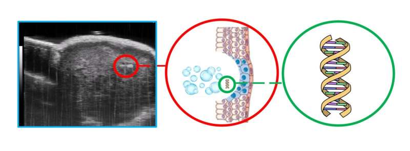
Ultrasound imaging offers a valuable and noninvasive way to find and monitor cancerous tumors. However, much of the most crucial information about a cancer, such as specific cell types and mutations, cannot be learned from imaging and requires invasive and damaging biopsies. One research group developed a way to employ ultrasound to extract this genetic information in a gentler way.
At the University of Alberta, a team led by Roger Zemp explored how intense ultrasound can release biological indicators of disease, or biomarkers, from cells . These biomarkers, like miRNA, mRNA, DNA, or other genetic mutations , can help identify different types of cancer and inform the subsequent therapy.
Zemp presents this work Monday, May 13, at 8:30 a.m. EDT as part of a joint meeting of the Acoustical Society of America and the Canadian Acoustical Association , running May 13–17 at the Shaw Center located in downtown Ottawa, Ontario, Canada.
"Ultrasound, at exposure levels higher than is used for imaging, can create tiny pores in cell membranes, which safely reseal," Zemp said. "This process is known as sonoporation. The pores formed due to sonoporation were previously used to get drugs into cells and tissues. In our case, we care about releasing the contents of cells for diagnostics."
The ultrasound releases biomarkers from the cells into the bloodstream, increasing their concentration to a level high enough for detection. Using this method, oncologists can detect cancer and monitor its progression or treatment without the need for painful biopsies. Instead, they can use blood samples , which are easier to procure and less expensive.
"Ultrasound can enhance the levels of these genetic and vesicle biomarkers in blood samples by over 100 times," said Zemp. "We were able to detect panels of tumor-specific mutations, and now epigenetic mutations that were not otherwise detectable in blood samples."
Not only was this approach successful at detecting biomarkers , but it also boasts a lower price compared to conventional testing.
"We've also found that we can conduct ultrasound-aided blood testing to look for circulating tumor cells in blood samples with single-cell sensitivity for the price of a COVID test," said Zemp. "This is significantly cheaper than the current methods, which cost about $10,000 per test."
The team also demonstrated the potential for applying intense ultrasound to liquefy small volumes of tissue for biomarker detection. The liquefied tissue can be retrieved from blood samples or through fine-needle syringes, a much more comfortable option compared to the damaging core-needle alternative.
More accessible techniques to identify cancer will not only allow for earlier detection and treatment but will also allow medical practitioners to be nimble in their approach. They can establish if certain therapies are working without the risks and expenses often associated with repeated biopsies.
"We hope that our ultrasound technologies will benefit patients by providing clinicians a new kind of molecular readout of cells and tissues with minimal discomfort," said Zemp.
Explore further
Feedback to editors

Study highlights need for cell-type-specific therapies in treatment of HIV

Psilocybin may reverse anorexia's cognitive rigidity

Variations in 'ancient' immune cells linked to patients' survival in cancer

Study suggests two copies of APOE4 gene behind up to 20% of Alzheimer's cases
May 12, 2024

Researchers show genetic variant common among Black Americans contributes to large cardiovascular disease burden

First person to receive a genetically modified pig kidney transplant dies nearly 2 months later

New vaccine could protect against coronaviruses that haven't even emerged yet
May 11, 2024

Study links organization of neurotypical brains to genes involved in autism and schizophrenia

Study traces an infectious language epidemic

Visual experiences unique to early infancy provide building blocks of human vision, study finds
May 10, 2024
Related Stories

Study suggests liquid biopsy could detect and monitor aggressive small-cell lung cancer
Apr 10, 2024

Study discusses liquid biopsy as a game changer for early lung cancer detection
Mar 20, 2024

Proteins in milk and blood could one day let doctors detect breast cancer earlier, and save lives
Mar 16, 2024

Non-invasive imaging technique could reduce need for repeat cancer surgeries
Apr 24, 2020

Research fuels advances in bile duct cancer care
Jan 15, 2024

Liquid biopsy: A new tool for identifying and monitoring cancer
Jan 25, 2024
Recommended for you

Using MRI, engineers have found a way to detect light deep in the brain

Unobtrusive, implantable device could deepen our understanding of behavioral responses

Flicker stimulation shines in clinical trial for epilepsy

Study points to personalized treatment opportunities for glioblastoma

Scientists make progress on new charged particle therapy for cancer
Let us know if there is a problem with our content.
Use this form if you have come across a typo, inaccuracy or would like to send an edit request for the content on this page. For general inquiries, please use our contact form . For general feedback, use the public comments section below (please adhere to guidelines ).
Please select the most appropriate category to facilitate processing of your request
Thank you for taking time to provide your feedback to the editors.
Your feedback is important to us. However, we do not guarantee individual replies due to the high volume of messages.
E-mail the story
Your email address is used only to let the recipient know who sent the email. Neither your address nor the recipient's address will be used for any other purpose. The information you enter will appear in your e-mail message and is not retained by Medical Xpress in any form.
Newsletter sign up
Get weekly and/or daily updates delivered to your inbox. You can unsubscribe at any time and we'll never share your details to third parties.
More information Privacy policy
Donate and enjoy an ad-free experience
We keep our content available to everyone. Consider supporting Science X's mission by getting a premium account.
E-mail newsletter
ORIGINAL RESEARCH article
Efficacy and safety of ultrasound-guided pulsed radiofrequency in the treatment of the ophthalmic branch of postherpetic trigeminal neuralgia.

- The Affiliated Hospital of Southwest Medical University, Luzhou, China
The final, formatted version of the article will be published soon.
Select one of your emails
You have multiple emails registered with Frontiers:
Notify me on publication
Please enter your email address:
If you already have an account, please login
You don't have a Frontiers account ? You can register here
Abstract Objective: To investigate the efficacy and safety of ultrasound-guided pulsed radiofrequency (PRF) targeting supraorbital nerve for treating the ophthalmic branch of postherpetic trigeminal neuralgia. Methods: A retrospective cohort study was conducted involving patients who presented at the Department of Pain, Affiliated Hospital of Southwest Medical University from January 2015 to January 2022. The patients were diagnosed with the first branch of postherpetic trigeminal neuralgia. In total, 63 patients were included based on the inclusion and exclusion criteria. The patients were divided into the following two groups based on the treatment method used: nerve block (NB) group (n = 32) and PRF + NB group (radiofrequency group, n = 31). VAS scores, PSQI scores, and dose of pregabalin taken before and after treatment were compared between the two groups. Results: The postoperative VAS score, PSQI score, and pregabalin dose were significantly decreased in both groups. Furthermore, significant differences were found between the two groups at each time point preoperative and postoperative (P < 0.05). After 6 months of treatment, the excellent rate of VAS score in the radiofrequency group was 70.96% and the overall effective rate was 90.32%, which was higher than that in the NB group. The difference in the efficacy was statistically significant (P < 0.05). Conclusion: PRF targeting the supraorbital nerve can effectively control the pain in the first branch of the trigeminal nerve after herpes, enhance sleep quality, and reduce the dose of pregabalin.
Keywords: Herpes Zoster, Pulsed radiofrequency, Trigeminal Neuralgia, Ultrasound guidanc e, Nerve Block
Received: 11 Mar 2024; Accepted: 13 May 2024.
Copyright: © 2024 LI, Gong, Zhang and Ou. This is an open-access article distributed under the terms of the Creative Commons Attribution License (CC BY) . The use, distribution or reproduction in other forums is permitted, provided the original author(s) or licensor are credited and that the original publication in this journal is cited, in accordance with accepted academic practice. No use, distribution or reproduction is permitted which does not comply with these terms.
* Correspondence: Fubo LI, The Affiliated Hospital of Southwest Medical University, Luzhou, China
Disclaimer: All claims expressed in this article are solely those of the authors and do not necessarily represent those of their affiliated organizations, or those of the publisher, the editors and the reviewers. Any product that may be evaluated in this article or claim that may be made by its manufacturer is not guaranteed or endorsed by the publisher.
- Open access
- Published: 09 May 2024
Correlation of alanine aminotransferase levels and a histological diagnosis of steatohepatitis with ultrasound-diagnosed metabolic-associated fatty liver disease in patients from a centre in Nigeria
- O. J. Kolawole 1 ,
- M. M. Oje 2 ,
- O. A. Betiku 3 ,
- O. Ijarotimi 4 ,
- O. Adekanle 4 &
- D. A. Ndububa 4
BMC Gastroenterology volume 24 , Article number: 147 ( 2024 ) Cite this article
124 Accesses
Metrics details
Metabolic-associated fatty liver disease (MAFLD) is defined as the occurrence of hepatic fat accumulation in patients with negligible alcohol consumption or any other cause of hepatic steatosis. This study aimed to correlate the ultrasound-based diagnosis of MAFLD with the histological diagnosis of nonalcoholic steatohepatitis (NASH) and alanine aminotransferase (ALT) levels in patients with MAFLD.
This was a hospital-based cross-sectional study of 71 patients with MAFLD diagnosed by ultrasound. Percutaneous liver biopsy was performed for histological evidence of NASH in all patients, regardless of liver function test (LFT) values, provided that they had no contraindications. Liver histology was graded using the NASH Clinical Research Network MAFLD Activity Score. The data obtained were entered into SPSS version 21 and analysed using descriptive and inferential statistics. The significance level was set at < 0.05.
A total of 71 patients (26 males and 45 females) with MAFLD were included. Thirty-nine (76.5%) patients with MAFLD and normal ALT levels had NASH, while 14 (82.4%) had elevated ALT levels. There was no statistically significant difference in the histological grade of NASH between patients with normal and elevated ALT levels. A weak correlation was found between the severity of steatosis on ultrasound scan and NASH incidence ( p = 0.026). The sensitivity and specificity of ALT levels for predicting NASH according to the area under the receiver operating characteristics (AUROC 0.590) at an ALT cut-off value of 27.5 IU/L were 55.8% and 64.7%, respectively.
NASH can occur in patients with MAFLD, irrespective of alanine transaminase (ALT) levels, and ultrasound grading of the severity of steatosis cannot accurately predict NASH. Liver biopsy remains the investigation of choice.
Peer Review reports
Introduction
Metabolic-associated fatty liver disease (MAFLD) is defined as the occurrence of hepatic fat accumulation after the exclusion of other causes of hepatic steatosis, such as liver disease and excessive alcohol consumption [ 1 ]. In clinical practice, MAFLD is regarded as the presence of fatty liver on ultrasonography in the absence of known secondary causes of fatty liver disease [ 2 ]. Nonalcoholic steatohepatitis (NASH) is a histologic category of MAFLD characterized by hepatic steatosis and inflammation with hepatocyte injury with or without fibrosis [ 3 ]. NASH can progress to cirrhosis, liver failure and hepatocellular carcinoma and is the third most common risk factor for hepatocellular carcinoma after viral hepatitis and alcohol consumption [ 2 ]. MAFLD is one of the most common liver diseases worldwide, and NASH may soon become the most common indication for liver transplantation [ 3 ]. MAFLD affects approximately 80 to 100 million individuals in the USA, among whom nearly 25% progress to NASH [ 1 ].
Liver biopsy is the gold standard for the diagnosis of MAFLD because it is the only way of assessing the presence of inflammation and fibrosis [ 4 ].
A study in Nigeria reported the prevalence of fatty liver to be 8.7% [ 5 ] and that of MAFLD to be 1.38% [ 6 ]. The increasing incidence of MAFLD because of increasing rates of obesity, type 2 DM and dyslipidaemia is worrisome. NASH is the most important component of the MAFLD spectrum, irrespective of the aminotransferase level. There is a paucity of literature on the indications for liver biopsy in patients with MAFLD with respect to their serum alanine aminotransferase (ALT) level to perform NASH assessment and provide prompt intervention. According to a population study in Sweden [ 7 ], once MAFLD progresses to NASH, cirrhosis survival decreases. The objectives of this study were therefore to determine the proportion of patients with MAFLD and normal ALT levels who have histological evidence of NASH and the proportion of patients with MAFLD and elevated ALT levels who have NASH; to assess the MAFLD activity score in patients with MAFLD and NASH; and to relate the histological findings to the ultrasound grading of fatty liver disease. Hence, there is a need for this study in Nigeria.
Materials and methods
This was a hospital-based cross-sectional study of 71 patients with MAFLD. The study period was from July 2019 to July 2021. The study was carried out in the gastroenterology clinic of a university teaching hospital in southwest Nigeria. The study included patients who were aged 18 years and older and had an ultrasound diagnosis of fatty liver disease and were either visiting the gastroenterology clinic or referred from the general outpatient, endocrinology and metabolic clinic or the cardiology clinic of the teaching hospital who met the inclusion criteria.
The inclusion criteria were as follows:
Individuals aged 18 years and older with an ultrasound diagnosis of fatty liver who were diagnosed by a specialist radiologist using the conventional USG system and graded according to fatty liver disease severity (grade 1: when liver echogenicity is just increased; grade 2: when the echogenic liver obscures the echogenic walls of the portal vein branches; and grade 3: when the echogenic liver obscures the diaphragmatic outline).
Patients with fatty liver disease and no significant alcohol consumption (≤ 21 units/week or < 30 g/day for men and ≤ 14 units/week or < 20 g/day for women) [ 1 , 8 ].
Hypertension in the study subjects was defined as a blood pressure ≥ 140/90 mmHg, which was checked one time, or on the use of antihypertensive drugs.
Diabetes status was defined according to the World Health Organization (WHO) guidelines [ 9 ] as follows:
A fasting plasma glucose level greater than or equal to 7 mmol/L (126 mg/dl).
A 2-hour postprandial plasma glucose level greater than or equal to 11.1 mmol/L (200 mg/dl).
Already receiving treatment for DM.
Obesity was defined as a body mass index ≥ 30 kg/m 2 , a waist circumference > 102 cm (40 inches) in men and > 88 cm (35 inches) in women and/or a waist/hip ratio > 0.90 in men and > 0.85 in women [ 10 ].
Dyslipidaemia was defined as having one or more of the following: an LDL-C level ≥ 100 mg/dl (2.6 mmol/l), a total cholesterol (TC) level ≥ 200 mg/dl (5.18 mmol/l), a triglyceride level ≥ 150 mg/dl (1.7 mmol/l) or a high-density lipoprotein cholesterol (HDL) level ≤ 40 mg/dl (1 mmol/l) in men and ≤ 50 mg/dl (1.3 mmol/l) in women [ 11 ].
Patients with the following conditions were excluded:
Patients with hepatitis B virus, hepatitis C virus or HIV infection.
Patients with a history of medication use, such as glucocorticoid and synthetic oestrogen, amiodarone, tamoxifen, methotrexate, tetracycline, antiretroviral drugs, or toxic mushrooms.
Patients with comorbidities such as congestive heart failure, chronic obstructive pulmonary disease, end-stage chronic kidney disease and malignancy of any origin.
Pregnant women.
The sample size was calculated using the formula for estimating single proportions and a prevalence of MAFLD (8.7%) [ 5 ] as follows: \({\rm{n = }}\frac{{{\rm{Z}}{{\rm{\alpha }}^{\rm{2}}}{\rm{P}}{{\rm{q}}^{{\rm{12}}}}}}{{{{\rm{d}}^{\rm{2}}}}}\) [ 12 ]. This resulted in a sample size of 71. Eligible patients were recruited consecutively until the sample size was met.
A structured proforma was used to obtain the following: sociodemographic information; data on the history of the present illness, alcohol intake, psychoactive substance use and drug use; risk factors for MAFLD (hypertension, type 2 diabetes mellitus, and obesity) and liver disease; and physical examination, laboratory investigation, ultrasonography, liver biopsy and final diagnosis data.
All patients who consented to participate in the study had basic laboratory test results, which included full blood count, viral hepatitis B and C, and clotting profile data in addition to liver function test data obtained using an agape kit and processed using an automated Cobas machine via a method comparable to the International Federation of Clinical Chemistry guide. All patients underwent percutaneous liver biopsy irrespective of liver function test results. Liver biopsies were prepared according to standard guidelines, stained with appropriate stains and read by a pathologist with a special interest in liver histology. Liver histological evidence of steatohepatitis (NASH) included steatosis of more than 5% hepatocytes, inflammatory cell infiltrates, ballooning degeneration with or without Mallory bodies, and peri-cellular/peri-venular fibrosis [ 1 ]. NASH was classified using the NASH CRN score (steatosis + ballooning + lobular inflammation). Patients with a NASH score of 5 and above were considered to have definite NASH, while those with a score of 1–4 were classified as not having NASH [ 13 ].
Ethical approval was obtained from the hospital Ethics and Research Committee under IRB/IEC/0004553 and NHREC/27/02/2009a and protocol number ERC/2019/06/12. Informed and written consent was obtained from all the participants after providing a detailed explanation of the purpose of the study, procedures, and liver biopsy. The data obtained were entered into the Statistical Package for Social Sciences version (SPSS) version 21. The data were analysed using descriptive and inferential statistics. The significance level was set at a p value < 0.05 and a 95% confidence interval.
A total of 71 patients were recruited and completed the study. The age range was 19 years to 69 years, with a mean age (SD) of 50.99 (± 11.76) years. There were more females than males (63.4% vs. 36.6%). The majority of the participants were of Yoruba ethnicity (93%), were married (90.14%) and practised Christianity (87.3%). More than half (44; 62%) of the participants had a tertiary education, and most of them were either civil servants (33; 46.5%) or traders (24; 33.8%) (Table 1 ).
Approximately 37 (52.1%) of the participants had symptoms, and among them,36 (50.7%) had right upper abdominal pain. The majority of the participants had hepatomegaly (51 [71.8%] patients) on abdominal examination. Only 11 (15.5%) patients were taking statins (Table 2 ).
The medical conditions of the patients were as follows: hypertension in 43 patients (60.6%), dyslipidaemia in 52 patients (73.2%) and type 2 DM or impaired fasting in 13 patients (18.3%). Twenty (76.9%) of the males and 42 (93.3%) of the females had central obesity. According to BMI, 37 (52.1%) of the participants were obese, of whom 3 (4.2%) and 28 (39.4%) were morbidly obese and overweight, respectively (Table 3 ).
Histology of the 71 liver biopsy samples revealed that 1 (1.4%) participant had a normal liver, 2 (2.8%) participants had fragmented liver tissue that could not be characterized using the NASH CRN score, 53 (74.6%) participants had NASH, and 15 (21.1%) participants did not have NASH (Fig. 1 ).
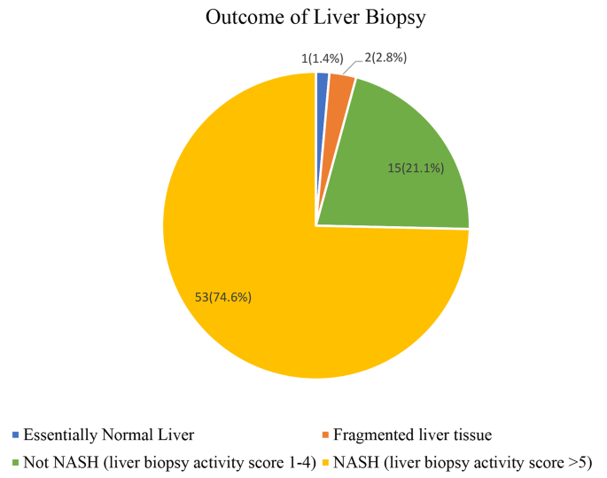
Outcome of liver biopsy
Using the NASH CRN score for liver biopsy grading, 53 participants (77.9%) with a CRN score of 5–8 had definite NASH, 11 participants (16.2%) with a CRN score of 3–4 had borderline NASH, and 4 participants (5.9%) with a score of 1–2 did not have NASH (Fig. 2 ).
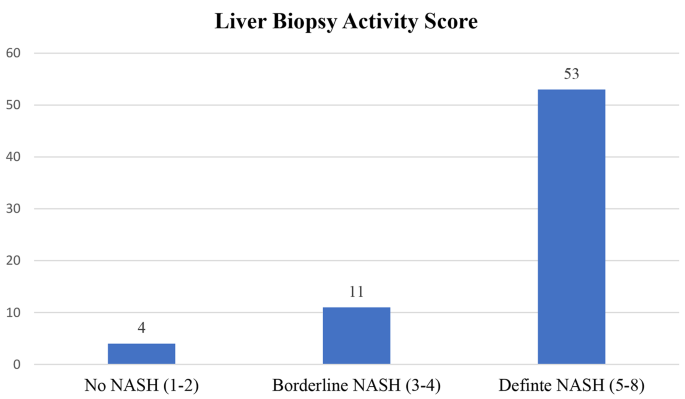
Frequency of NASH-CRN scores among the study participants
According to the ALT values, 51 patients had normal ALT levels (< 40 IU/L); among these patients, 39 (76.5%) had NASH, and 12 (23.5%) did not. Similarly, among the 17 patients with MAFLD with elevated ALT levels (> 40 IU/L), 14 (82.4%) had NASH, while 3 (17.6%) did not (Table 4 ).
Patients with higher grades of fatty liver disease on abdominal ultrasound scan also had a greater incidence of NASH; 61.9% of patients with grade 1 steatosis on ultrasound had NASH; 84.1% of patients with grade 2 steatosis on ultrasound had NASH; and 100% of patients with grade 3 steatosis on ultrasound had NASH. Additionally, patients with definite NASH according to histology had higher proportions of Grade 1, 2 and 3 fatty liver disease according to ultrasound (61.9%, 84.1% and 100%, respectively), while the proportions of patients with steatosis and without NASH according to ultrasound were as follows: Grade 1 (38.1%), Grade 2 (15.9%) and Grade 3 (0%). There was a weak correlation between the grade of fatty liver disease determined by ultrasound and NASH incidence ( p = 0.027) (Tables 5 & 6 ).
The ROC curves of the NASH CRN score and serum ALT concentration showed that the area under the curve (diagnostic accuracy) was 0.590 ( p = 0.268, 95% CI = 0.435–0.745). At an ALT cut-off value of 27.5 IU/L, the sensitivity and specificity were 55.8% and 64.7%, respectively, with positive and negative predictive values of 82.9% and 32.4% (Fig. 3 ; Table 7 ).
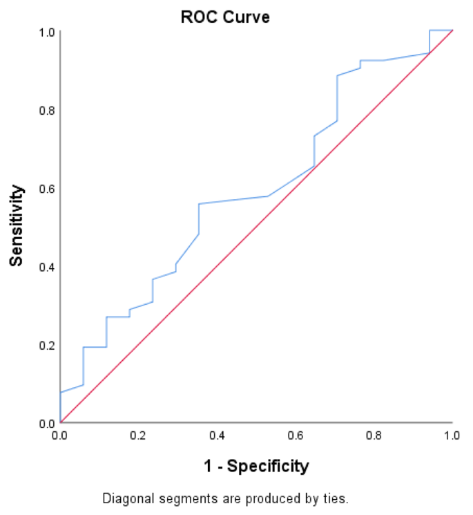
ROC curves for the NASH CRN score and serum ALT concentration
The majority of the patients with MAFLD were females, with a female-to-male ratio of 1.73:1. Studies including premenopausal women have reported male-dominated findings, while in studies including postmenopausal women, the effect of hormonal protection was lost; therefore, sex differences may be the same, or female MAFLD patients may dominate [ 1 ]. Most women in this study had reached menopause, with a mean age of 49.93 ± 12 years, which may explain why females predominated in this study. Additionally, there was no significant difference between the mean age of the participants with normal and elevated ALT levels or between those with and without NASH. These findings are consistent with the findings of Uslusoy et al. [ 14 ].
This study revealed that the overall risk factors for MAFLD were hypertension, type 2 DM, dyslipidaemia, obesity, and impaired fasting glucose. Moreover, more patients with NASH had these conditions than did those without NASH; these findings are similar to previous reports [ 15 , 16 , 17 , 18 ].
The most widely used imaging test for hepatic steatosis is ultrasound because it is readily available, inexpensive, and safe [ 2 ]. The majority of the patients in this study had grade 2 hepatic steatosis on ultrasound. This study revealed that the greater the grade of steatosis on ultrasound was, the greater the proportion of participants with definite NASH. There is a paucity of data in the literature concerning the ability of ultrasound to distinguish between simple steatosis and histological features of NASH. Saadeh et al. [ 19 ] previously reported that none of the radiological modalities could detect hepatocyte features important for diagnosing NASH. This study further corroborated the findings of Saadeh et al. [ 19 ] However, there was no statistically significant relationship between USS for hepatic steatosis and NASH incidence. A statistically weak relationship was observed between the grade of hepatic steatosis determined by ultrasound and the incidence of NASH. Similar findings were reported by Abdel et al. [ 20 ] Therefore, this study showed that there was a significant correlation between the USS grade and the presence of NASH and the NASH CRN score.
ALT levels were measured using a cut-off value of 40 IU/L, which is the standard laboratory reference value based on the kit that was used in the automated Cobas machine and is comparable to the International Federation of Clinical Chemistry Method. Three quarters (75%) of the participants had normal ALT levels (< 40 IU/L), while one quarter (25%) had elevated ALT levels (> 40 IU/L). Ma et al. [ 21 ] reported that only 25% of the patients had normal ALT levels in a systematic review and meta-analysis. This study showed that the ALT level does not reflect what occurs in the liver of a patient with MAFLD, which is consistent with earlier reports [ 21 , 22 , 23 ]. These findings may be explained by the findings of Ma et al. [ 21 ], who showed that a normal ALT level is closely associated with diabetes, metabolic syndrome and steatosis grade 1 compared with steatosis grades 2 and 3. It was also associated with sex, and females with NAFLD tended to have normal ALT levels.
This study also showed that more patients with normal ALT levels had simple steatosis than did those with elevated ALT levels. Wong et al. [ 18 ] and Khosravi et al. [ 24 ] found that ALT levels did not correlate with histological parameters in patients with MAFLD. Wong et al. [ 15 ] reported that NASH and significant fibrosis can be found in participants with normal ALT levels. Another multicentre study involving 733 patients with MAFLD who underwent liver biopsy reported that advanced fibrosis did not correlate with alanine transaminase (ALT) levels [ 25 ]. This study revealed variability in ALT levels and the incidence of NASH using the NASH CRN score. The prevalence of NASH did not differ between patients with normal ALT levels and those with elevated ALT levels. Similarly, other authors have reported varying percentages of patients with NASH and normal ALT levels and MAFLD [ 26 , 27 , 28 ].
This study revealed significant differences in BMI, systolic blood pressure and AST levels between patients with and without NASH and between patients with and without elevated ALT levels. These findings suggested that patients with MAFLD who are obese and hypertensive with elevated ALT and AST levels are at increased risk of progression to NASH.
This study revealed that the frequency of biopsy-confirmed NASH was highly comparable to that of another report by Ajmera et al. [ 29 ] A meta-analysis by Dufour et al. [ 30 ] reported a wide range of (15.9–68.3%) of patients with MAFLD who underwent liver biopsy and were confirmed to have NASH.
There is no specific cut-off value for ALT in patients with MAFLD. The area under the receiver operating curve (AUROC) for ALT levels relating to the incidence of NASH was 0.590. At an ALT cut-off value of 27.5 IU/L, the sensitivity and specificity were 55.8% and 64.7%, respectively. The study failed to demonstrate an optimal ALT level that would mostly predict the incidence of NASH. These findings are comparable with those of other studies of patients with MAFLD [ 18 , 28 ].
Limitations of the study
The intake of alcohol was estimated by patient recall, which may not be accurate.
The sample size of the study was small, and a larger sample size would have increased the power of the study.
This was a cross-sectional study, and patients were not followed up to determine what would have become of those without NASH; it is possible that some may have progressed to NASH.
The ultrasonography technique used to detect hepatic fat in this study had poor sensitivity for diagnosing steatosis when it was less than 30%. A more advanced test, such as magnetic resonance imaging (MRI), including magnetic resonance spectroscopy, would have been a better option because it can detect the presence of hepatic fat greater than 5.6%, which is the defining threshold with an accuracy of 100%.
Strength of the study
Liver biopsy and histology were performed for all patients with MAFLD irrespective of their serum ALT levels. This approach is the gold standard for diagnosing NASH and detecting early fibrosis, cirrhosis, and hepatocellular carcinoma.
The proportion of patients with NASH and normal ALT levels was greater than that of patients with NASH and elevated ALT levels. The variability in ALT levels and the histological diagnosis of NASH showed that the ALT level is not a reliable marker for diagnosing NASH. In addition, the sensitivity and specificity of the ALT level for predicting NASH were found to be low. There is no ideal cut-off value for ALT levels using the AUROC for predicting the incidence of NASH. Thus, liver biopsy may be necessary for all patients with MAFLD, irrespective of their ALT level, to detect the presence of NASH.
This study also showed that despite the increasing incidence of NASH with increasing steatosis grade on ultrasound, there was a weak correlation between the grade of steatosis on ultrasound and the incidence of NASH.
Data availability
The datasets used and/or analysed during the current study available from the corresponding author on reasonable request.
Abbreviations
Alanine Aminotransferase
Aspartate Aminotransferase
Area under receivers operating curve
Body Mass Index
Clinical Research Network
Diabetes mellitus
Type 2 Diabetes mellitus
High density lipoprotein cholesterol
Human Immunodeficiency Virus
Low density lipoprotein cholesterol
Liver function test
Magnetic Resonance Imaging
Metabolic syndrome
Metabolic associated Fatty Liver Disease
Non-alcoholic fatty liver disease
Non-alcoholic Steatohepatitis
National Health and Nutrition Examination Survey
National Health Research Ethics committee
Obafemi Awolowo University Teaching Hospital Complex
Receiver operating curve
Standard deviation
Statistical package for the social sciences
Total Cholesterol
United States of America
Ultrasonography
Ultrasound scan
World Health Organisation
Perumpail BJ, Khan MA, Yoo ER, Cholankeril G, Kim D, Ahmed A. Clinical epidemiology and disease burden of nonalcoholic fatty liver disease. World J Gastroenterol. 2017;23(47):8263–76.
Article PubMed PubMed Central Google Scholar
Onyekwere CA, Ogbera AO, Samaila AA, Balogun BO, Abdulkareem FB. Nonalcoholic fatty liver disease: Synopsis of current developments. Niger J Clin Pract. 2015;18(6):703–12.
Article CAS PubMed Google Scholar
Chalasani N, Younossi Z, Lavine JE, Diehl AM, Brunt EM, Cusi K, et al. AASLD practice guideline; the diagnosis and management of non-alcoholic fatty liver disease: Practice Guideline by the American Association for the study of Liver diseases. American College of Gastroenterology, and the American Gastroenterological Assoc; 2012. pp. 2005–23.
Chrysoula B, Petros L. No n-alcoholic fatty liver disease vs non alcoholic steatohepatitis: pathological and clinical implications. Curr Vasc Pharmacol. 2018;16(3):214–8.
Article Google Scholar
Onyekwere CA, Ogbera AO, Balogun BO. Nonalcoholic fatty liver disease and the metabolic syndrome in an urban hospital serving an African community. Ann Hepatol. 2011;10(2):119–24.
Article PubMed Google Scholar
Ndububa DA, Ojo OS, Adetiloye VA, Aladegbaiye AO, Adebayo RA, Adekanle O. The contribution of alcohol to chronic liver disease in patients from south-west Nigeria. Niger J Clin Pract. 2010;13(4):360–4.
CAS PubMed Google Scholar
Ekstedt M, Franzén LE, Mathiesen UL, Thorelius L, Holmqvist M, Bodemar Gör, Kechagias S. Long-term follow-up of patients with NAFLD and elevated liver enzymes. Hepatology. 2006;4(44):865–73.
Arab JP, Arrese M, Trauner M. Recent insights into the pathogenesis of nonalcoholic fatty liver disease. Annu Rev Pathol Mech Dis. 2018;13(1):321–50.
Article CAS Google Scholar
Report of a WHO/IDF Consultation. Definition and diagnosis of diabetes mellitus and intermediate hyperglycemia. In: world health organization. 2016. page 1.
Waist Circumference and Waist-Hip Ratio Report of a WHO Expert Consultation, Geneva. 2008;(December):8–11.
Grundy SM, Cleeman JI, Bairey Merz CN, Brewer HB, Clark LT, Hunninghake DB, et al. Implications of recent clinical trials for the National Cholesterol Education Program Adult Treatment Panel III guidelines. Circulation. 2004;110(2):227–39.
Daniel W. Biostastistics: A Foundation for analysis in the health sciences. Wiley New York. 1999;(7):141–2.
Brunt EM, Kleiner DE, Wilson LA, Belt P, Neuschwander-Tetri BA. Nonalcoholic fatty liver disease (NAFLD) activity score and the histopathologic diagnosis in NAFLD: distinct clinicopathologic meanings. Hepatology. 2011;53(3):810–20.
Uslusoy HS, Nak SG, Gülten M, Biyikli Z. Nonalcoholic steatohepatitis with normal aminotransferase values. World J Gastroenterol. 2009;15(15):1863–8.
Article CAS PubMed PubMed Central Google Scholar
Younossi ZM. Nonalcoholic fatty liver disease – a global public health perspective. J Hepatol. 2019;70(3):531–44.
Younossi ZM, Koenig AB, Abdelatif D, Fazel Y, Henry L, Wymer M. Global epidemiology of nonalcoholic fatty liver disease—Meta-analytic assessment of prevalence, incidence, and outcomes. Hepatology. 2016;64(1):73–84.
Younossi Z, Anstee QM, Marietti M, Hardy T, Henry L, Eslam M, et al. Global burden of NAFLD and NASH: Trends, predictions, risk factors and prevention. Nat Rev Gastroenterol Hepatol. 2018;15(1):11–20.
Wong VW, Wong GL, Tsang SW, Hui AY, Chan AW. Metabolic and histological features of non-alcoholic fatty liver disease patients with different serum alanine aminotransferase levels. Aliment Pharmacol Ther. 2009;29(11):387–96.
Sherif S, Zobair M, Erick M, Terry G, Janus P. The utility of Radiological Imaging in nonalcoholic fatty liver disease. Gastroenter. 2002;123:745–50.
Rahman A, Manasra A, Bani M. Correlation between ultrasound and histologic findings of fatty liver changes among non-alcoholic obese patients. J Med J. 2021;55(2):1–12.
Google Scholar
Ma X, Liu S, Zhang J, Dong M, Wang Y, Wang M. Proportion of NAFLD patients with normal ALT value in overall NAFLD patients: a systematic review and meta-analysis. BMC Gastroenterol. 2020;20(10):1–8.
Kunde SS, Lazenby AJ, Clements RHAG. Spectrum of NAFLD and diagnostic implications of the proposed new normal range for serum ALT in obese women. Hepatology. 2005;42:650–6.
Amarapurkar DN. Clinical spectrum and natural history of non-alcoholic steatohepatitis with normal alanine aminotransferase values. Trop Gastroenterol. 2004;25:130–4.
PubMed Google Scholar
Khosravi S, Alavian SM, Zare A, Daryani NE, Fereshtehnejad SM, Daryani NE, et al. Nonalcoholic fatty liver disease and correlation of serum alanin aminotransferase level with histopathologic findings. Hepat Mon. 2011;11(6):452–8.
PubMed PubMed Central Google Scholar
Angulo P, Hui JM, Marchesini G, Bugianesi E, George J, Farrell GC, et al. The NAFLD fibrosis score: a noninvasive system that identifies liver fibrosis in patients with NAFLD. Hepatology. 2007;45(4):846–54.
Fracanzani AL, Valenti L, Bugianesi E, Andreoletti M, Colli A, Vanni E, et al. Risk of severe liver disease in nonalcoholic fatty liver disease with normal aminotransferase levels: a role for insulin resistance and diabetes. Hepatology. 2008;48:792–8.
Lee JY, Kim KM. Prevalence and risk factors of non-alcoholic fatty liver disease in potential living liver donors in Korea: a review of 589 consecutive liver biopsies in a single center. J Hepatol. 2007;47:239–44.
Verma S, Jensen D, Hart J, Mohanty SR. Predictive value of ALT levels for non-alcoholic steatohepatitis (NASH) and advanced fibrosis in non-alcoholic fatty liver disease (NAFLD). Liver int. 2013;13(8):1398–405.
Ajmera V, Perito ER, Bass NM, Terrault NA, Yates KP, Gill R, et al. Novel plasma biomarkers associated with liver disease severity in adults with nonalcoholic fatty liver disease. Hepatology. 2017;65(1):65–77.
Dufour J-F, Scherer R, Balp M-M, McKenna SJ, Janssens N, Lopez P, et al. The global epidemiology of nonalcoholic steatohepatitis (NASH) and associated risk factors–A targeted literature review. Endocr Metab Sci. 2021;3(December 2020):100089.
Download references
Acknowledgements
1. Dr A.D Omisore, Department of Radiology, Obafemi Awolowo University Teaching Hospital Complex, Ile-Ife, Osun State Nigeria for her professional support during this study. 2. Dr S.A Olowookere, Obafemi Awolowo University Ile-Ife, Osun State Nigeria for his professional advise. 3. Mr Olorunyomi Kolawole, Thornford Park Hospital, Thatcham England for his technical help and writing assistance.
Not applicable.
Author information
Authors and affiliations.
Department of General Medicine, Frimley Health NHS Foundation Trust, Wexham Park Hospital, Slough, SL2 4HL, UK
O. J. Kolawole
Department of Medicine, LAUTECH Teaching Hospital, Ogbomoso, Oyo State, Nigeria
Department of Morbid Anatomy, College of Health Sciences, Obafemi Awolowo University Teaching Hospital Complex, Ile-Ife, Osun State, Nigeria
O. A. Betiku
Department of Medicine, Faculty of Clinical Sciences, Obafemi Awolowo University and Obafemi Awolowo University Teaching Hospital Complex, Ile-Ife, Osun State, Nigeria
O. Ijarotimi, O. Adekanle & D. A. Ndububa
You can also search for this author in PubMed Google Scholar
Contributions
1. OJ, O Adekanle, O Ijarotimi and DA - selection of research Topic. 2. OJ, MM, O Ijarotimi, and O Adekanle - data collection and recruitement of participants. 3. OA Betiku- reviewed histology slides. 4. OJ, MM, O Adekanle and DA contributed to analysis of data. 5. OJ, O Adekanle wrote the main manuscript text. All authors reviewed the manuscript.
Corresponding author
Correspondence to O. J. Kolawole .
Ethics declarations
Ethics approval and consent to participate.
Ethical approval was obtained from the Ethics and Research Committee of the Obafemi Awolowo University Teaching Hospital Ile-Ife, Nigeria where the study was carried out under IRB/IEC/0004553 and NHREC/27/02/2009a and protocol number ERC/2019/06/12. The Ethics committee of the hospital is under the National Health Research Ethics committee (NHREC) of the Federal ministry of Health in Nigeria which is affiliated to the West African Bio-ethics centre. Informed and written consent was obtained from all the participants after providing a detailed explanation of the purpose of the study, procedures, and liver biopsy.
Consent for publication
Consent was taken from each participant for the research.
Competing interests
The authors declare no competing interests.
Additional information
Publisher’s note.
Springer Nature remains neutral with regard to jurisdictional claims in published maps and institutional affiliations.
Rights and permissions
Open Access This article is licensed under a Creative Commons Attribution 4.0 International License, which permits use, sharing, adaptation, distribution and reproduction in any medium or format, as long as you give appropriate credit to the original author(s) and the source, provide a link to the Creative Commons licence, and indicate if changes were made. The images or other third party material in this article are included in the article’s Creative Commons licence, unless indicated otherwise in a credit line to the material. If material is not included in the article’s Creative Commons licence and your intended use is not permitted by statutory regulation or exceeds the permitted use, you will need to obtain permission directly from the copyright holder. To view a copy of this licence, visit http://creativecommons.org/licenses/by/4.0/ . The Creative Commons Public Domain Dedication waiver ( http://creativecommons.org/publicdomain/zero/1.0/ ) applies to the data made available in this article, unless otherwise stated in a credit line to the data.
Reprints and permissions
About this article
Cite this article.
Kolawole, O.J., Oje, M.M., Betiku, O.A. et al. Correlation of alanine aminotransferase levels and a histological diagnosis of steatohepatitis with ultrasound-diagnosed metabolic-associated fatty liver disease in patients from a centre in Nigeria. BMC Gastroenterol 24 , 147 (2024). https://doi.org/10.1186/s12876-024-03237-4
Download citation
Received : 28 December 2023
Accepted : 22 April 2024
Published : 09 May 2024
DOI : https://doi.org/10.1186/s12876-024-03237-4
Share this article
Anyone you share the following link with will be able to read this content:
Sorry, a shareable link is not currently available for this article.
Provided by the Springer Nature SharedIt content-sharing initiative
- Liver biopsy
- Alanine aminotransferase
BMC Gastroenterology
ISSN: 1471-230X
- Submission enquiries: [email protected]
- General enquiries: [email protected]
Cornell Chronicle
- Architecture & Design
- Arts & Humanities
- Business, Economics & Entrepreneurship
- Computing & Information Sciences
- Energy, Environment & Sustainability
- Food & Agriculture
- Global Reach
- Health, Nutrition & Medicine
- Law, Government & Public Policy
- Life Sciences & Veterinary Medicine
- Physical Sciences & Engineering
- Social & Behavioral Sciences
- Coronavirus
- News & Events
- Public Engagement
- New York City
- Photos of the Week
- Big Red Sports
- Freedom of Expression
- Student Life
- University Statements
- Around Cornell
- All Stories
- In the News
- Expert Quotes
- Cornellians
Ultrasound experiment identifies new superconductor
By kate blackwood college of arts and sciences.
With pulses of sound through tiny speakers, Cornell physics researchers have clarified the basic nature of a new superconductor.
Since it was found to be a superconductor about five years ago, uranium ditelluride has created a lot of buzz in the quantum materials community – and a lot of confusion, with more than a dozen theories about the true nature of its superconducting properties. Some suggested valuable possibilities for quantum computing.
In an experiment, Brad Ramshaw , associate professor of physics in the College of Arts and Sciences (A&S) and colleagues used ultrasound to gather direct evidence that uranium ditelluride has a single-component superconducting order parameter, ruling out a more exotic type of superconductor that would have been exciting news for quantum computing. But setting a baseline of data for the material’s intrinsic superconductivity still leaves the door open for discovering additional complex possibilities through further study.
The experiment establishes that recent technical developments in the Ramshaw lab make pulse-echo ultrasound, which uses sound pulses to examine the mechanical stiffness of quantum materials, a trustworthy and desirable technique for examining superconducting materials.
“ Single-component Superconductivity in UTe2 at Ambient Pressure ” was published in Nature Physics on May 9. Ramshaw is corresponding author with doctoral student Florian Theuss as first author. Doctoral student Avi Shragai and former postdoctoral researcher Gael Grissonnanche, now faculty at the Institut Polytechnique in Paris, contributed, along with collaborators from the University of Maryland and the University of Wisconsin, Milwaukee.
“All superconductors have zero resistance, but at a subtler level, there are different flavors of superconductors,” Ramshaw said. “Researchers are interested in finding these different flavors because, one, we don’t even know if they exist, even though we know in theory that they can exist. And two, they can be used in technologies like quantum computation. You need new types of superconductors for new technologies.”
A strange combination of properties in uranium ditelluride suggested at first that it could be this new type of superconductor. Its critical temperature – how cold it has to get before transitioning into a superconducting state – is relatively low, about 2 kelvin. But its low critical temperature is paired with a very high critical field – the measure of how much magnetic field it can withstand before the superconducting state collapses.
“We would normally expect it to withstand one or two tesla, but it can withstand around 60,” Ramshaw said. It is nearly 100 times stronger than any magnetic field you’d encounter in everyday life. That tells us there’s something weird, that maybe it’s one of those new flavors of superconductivity.”
Ramshaw and his collaborators wanted to find whether the material has, as some theories and existing experiments predicted, a multi-component superconducting order parameter, entailing exotic effects, or single-component order parameter, still potentially exotic but much more constrained.
Theuss led an experiment using pulse-echo ultrasound on a 1 millimeter by 1 millimeter sample to uncover the interplay between the structure and superconductivity in uranium ditelluride. The technique measures the speed of a sound pulse moving through a material, the same principle as medical ultrasound imaging. The difference is that instead of producing images, the researchers measured the sound speed to detect the change in stiffness of the material as it cooled down to and past the critical temperature.
“We can measure the distance between the sound echoes with phenomenal precision. That’s the real power of the experiment,” Ramshaw said.
Tiny speakers (transducers) attached to the sample pumped a sound pulse directly into the material in three different directions, measuring both three compression waves and three shear waves – the side-to-side vibrations only present in solids.
At the critical temperature, compression waves showed a sudden drop where the speed of sound plummeted, as expected for all superconductors. However, shear waves showed no such drop.
“If it was one of the exotics types of superconductivity people were proposing, these shear waves would have also had a drop,” Ramshaw said.
The researchers provide direct evidence that this material has a single-component order parameter. This conclusion, however, does not quell the excitement about superconductivity in uranium ditelluride, which has many interesting aspects worth further study, including its extraordinarily strong repulsion to magnetism.
Applying pressure or magnetic fields below the critical temperature could change the type of superconductivity, perhaps even creating the elusive two-component spin-triplet superconductivity, Ramshaw said. The current study provides a data-based place to begin.
“Definitely there’s more to come in this material. We’ve only just started,” said Theuss, who has worked with uranium ditelluride for much of his Ph.D. candidacy. “But if you want to explain these complicated things, you have to start at the basic intrinsic facts of the superconductivity in Ute2.”
Funding for this research comes from the Office of Basic Energy Sciences of the United States Department of Energy; the Cornell Center for Materials Research Shared Facilities which are supported through the National Science Foundation. The University of Maryland researchers received support from the Gordon and Betty Moore Foundation; the Maryland Quantum Materials Center; and the National Institute of Standards and Technology.
Kate Blackwood is a writer for the College of Arts and Sciences.
Media Contact
Becka bowyer.
Get Cornell news delivered right to your inbox.
You might also like

Gallery Heading
Lab on a Chip
A vibrating capillary for ultrasound rotation manipulation of zebrafish larvae †.

* Corresponding authors
a Acoustic Robotics Systems Laboratory, Institute of Robotics and Intelligent Systems, Department of Mechanical and Process Engineering, ETH Zurich, Säumerstrasse 4, CH-8803 Zurich, Switzerland E-mail: [email protected]
b Neuhauss Laboratory, Department of Molecular Life Sciences, University of Zurich, Winterthurerstrasse 190, CH-8057 Zurich, Switzerland
Multifunctional micromanipulation systems have garnered significant attention due to the growing interest in biological and medical research involving model organisms like zebrafish ( Danio rerio ). Here, we report a novel acoustofluidic rotational micromanipulation system that offers rapid trapping, high-speed rotation, multi-angle imaging, and 3D model reconstruction of zebrafish larvae. An ultrasound-activated oscillatory glass capillary is used to trap and rotate a zebrafish larva. Simulation and experimental results demonstrate that both the vibrating mode and geometric placement of the capillary contribute to the developed polarized vortices along the long axis of the capillary. Given its capacities for easy-to-operate, stable rotation, avoiding overheating, and high-throughput manipulation, our system poses the potential to accelerate zebrafish-directed biomedical research.

Supplementary files
- Supplementary information PDF (5171K)
- Supplementary movie MP4 (432K)
- Supplementary movie MP4 (2152K)
- Supplementary movie MP4 (1250K)
- Supplementary movie MP4 (1947K)
- Supplementary movie MP4 (5101K)
- Supplementary movie MP4 (15032K)
- Supplementary movie MP4 (1481K)
- Supplementary movie MP4 (959K)
Article information
Download Citation
Permissions.
A vibrating capillary for ultrasound rotation manipulation of zebrafish larvae
Z. Zhang, Y. Cao, S. Caviglia, P. Agrawal, S. C. F. Neuhauss and D. Ahmed, Lab Chip , 2024, 24 , 764 DOI: 10.1039/D3LC00817G
This article is licensed under a Creative Commons Attribution-NonCommercial 3.0 Unported Licence . You can use material from this article in other publications, without requesting further permission from the RSC, provided that the correct acknowledgement is given and it is not used for commercial purposes.
To request permission to reproduce material from this article in a commercial publication , please go to the Copyright Clearance Center request page .
If you are an author contributing to an RSC publication, you do not need to request permission provided correct acknowledgement is given.
If you are the author of this article, you do not need to request permission to reproduce figures and diagrams provided correct acknowledgement is given. If you want to reproduce the whole article in a third-party commercial publication (excluding your thesis/dissertation for which permission is not required) please go to the Copyright Clearance Center request page .
Read more about how to correctly acknowledge RSC content .
Social activity
Search articles by author, advertisements.

COMMENTS
Ultrasound articles from across Nature Portfolio. Atom. RSS Feed. Ultrasound is a non-invasive imaging technique that uses the differential reflectance of acoustic waves at ultrasonic frequencies ...
Correction: Feasibility of using a handheld ultrasound device to detect and characterize shunt and deep vein thrombosis in patients with COVID-19: an observational study. Authors: Rajkumar Rajendram, Arif Hussain, Naveed Mahmood and Mubashar Kharal. Citation:The Ultrasound Journal 2023 15 :44.
106 Ultrasound Essay Topic Ideas & Examples. Ultrasound technology has revolutionized the field of medicine, allowing healthcare professionals to visualize internal structures and organs without invasive procedures. As a result, ultrasound has become an essential tool for diagnosing and monitoring various medical conditions.
Medical ultrasound represented by ultrasound imaging is generally believed to be well-known area in research, because ultrasound imaging systems has been widely distributed and actively used in the world since the 1960s. ... Hence, this special issue is designed to bring in various ultrasound-related topics in medicine. We also tried to invite ...
The ongoing rapid expansion of point-of-care ultrasound ... This timely paper 10 aims to improve CCE research data reporting, such that clinical providers may more easily use data to support their clinical decision making when caring for critically ill patients. A critical appraisal of the existing literature from 2000-2017 was conducted, and ...
The Ultrasound Journal is an international, peer-reviewed journal designed for clinicians using point-of-care ultrasound in any environment or setting.The Ultrasound Journal is the official publication of WINFOCUS (World Interactive Network Focused on Critical UltraSound), the world's leading scientific organization committed to developing point-of-care ultrasound practice, research, education ...
Jan. 31, 2023 — New research found that using focused-ultrasound-mediated liquid biopsy in a mouse model released more tau proteins and another biomarker into the blood than without the ...
A wearable ultrasonic device to image cardiac function. Researchers have engineered a wearable device that adheres to the skin and uses ultrasound imaging and a deep learning model to produce a ...
The Journal of the British Medical Ultrasound Society, Ultrasound is dedicated solely to publishing ultrasound-related topics for users of this rapidly evolving specialty. It provides a forum for presentation of relevant scientific and technical advances in both diagnostic and therapeutic applications of ultrasound. View full journal description.
Ultrasound Quarterly provides coverage of the newest, most sophisticated ultrasound techniques as well as in-depth analysis of important developments in this dynamic field. The journal publishes reviews of a wide variety of topics including trans-vaginal ultrasonography, detection of fetal anomalies, color Doppler flow imaging, pediatric ultrasonography, and breast sonography.
The purpose of this editorial update is to provide readers with some recent information and to share my views on the journal and its future directions. During the last 4 years, Ultrasonography has published a total of 160 peer-reviewed articles. In 2017, the Ultrasonography home page had over 85,000 hits per month, with over 2,300 downloads of ...
Ultrasound Imaging - Current Topics presents complex and current topics in ultrasound imaging in a simplified format. It is easy to read and exemplifies the range of experiences of each contributing author. ... Open Access is an initiative that aims to make scientific research freely available to all. To date our community has made over 100 ...
The Journal for Vascular Ultrasound (JVU) is the official quarterly Journal of the Society for Vascular Ultrasound and publishes peer-reviewed articles on all aspects of the noninvasive diagnosis of vascular disease, including original research, reviews, case reports, letters to the editor, and editorials. Continuing Medical Education (CME) test questions are included with many JVU articles ...
Relationship Between Ultrasound Viewing and Proceeding to Abortion. Transrectal Ultrasound of the Prostate With a Biopsy. Iron-Based Catalysts Used in Water Treatment Assisted by Ultrasound. 88 Tourism Management Essay Topic Ideas & Examples 64 Virtual Team Essay Topic Ideas & Examples.
Keywords: medical ultrasound imaging, therapeutic ultrasound, non-destructive evaluation, nonlinear ultrasonics, physics of ultrasound, underwater ultrasound, ultrasonic transducers . Important Note: All contributions to this Research Topic must be within the scope of the section and journal to which they are submitted, as defined in their mission statements.
Transparent ultrasound chip improves cell stimulation and imaging. Ultrasound scans, best known for monitoring pregnancies or imaging organs, can also be used to stimulate cells and direct cell function. A team of Penn State researchers has developed an easier, more effective way to harness the technology for biomedical applications.
A thesis or dissertation, as some people would like to call it, is an integral part of the Radiology curriculum, be it MD, DNB, or DMRD. We have tried to aggregate radiology thesis topics from various sources for reference. Not everyone is interested in research, and writing a Radiology thesis can be daunting.
One of the most common uses of ultrasound is during pregnancy, to monitor the growth and development of the fetus, but there are many other uses, including imaging the heart, blood vessels, eyes, thyroid, brain, breast, abdominal organs, skin, and muscles. Ultrasound images are displayed in either 2D, 3D, or 4D (which is 3D in motion).
About this Research Topic. Submission closed. Musculoskeletal ultrasound is becoming an important and effective tool for the diagnosis of various soft tissue and joint disorders. It is accurate, safe, affordable, efficient, and dynamic. Musculoskeletal ultrasound is also a useful tool for guided therapy. The advances of musculoskeletal imaging ...
List of topics. Topics for thesis and projects are given below. Most of the topics can be adjusted to the students qualifications and wishes. Don't hesitate to take contact with the corresponding supervisor - we're looking forward to a discussion with you!
The goals of research in ultrasound usage in space environments are: (1) Determine accuracy of ultrasound in novel clinical conditions. (2) Determine optimal training methodologies, (3) Determine microgravity associated changes and (4) Develop intuitive ultrasound catalog to enhance autonomous medical care.
Topics in emergency abdominal ultrasonography Research. Edited by Luca Brunese and Antonio Pinto. Publication of this suppement has been funded by the University of Molise, Universiy of Siena, University of Cagliari, University of Ferrara and University of Turin. The Supplement Editors declare that they have no competing interests.
This project was funded by National Key Research and Development Program of China (No. 2023YFC3603803), TCL Charity Foundation Young Scholars Project (No. 2024001), Guangdong Basic and Applied ...
Background: Evidence suggests the plantar fascia and its interphase with the flexor digitorum brevis muscle can play a relevant role in plantar heel pain. Needling interventions could offer an appropriate treatment strategy to addressing this interface. Objective: We compared the accuracy and safety of ultrasound-guided versus palpation-guided procedures for the proper targeting of the ...
We trust that you will find a few clinical pearls or reminders that you could apply to your patients that you care for in your emergency department or other health setting. Search by keywords; disease process; condition; eponym or clinical features…. Ultrasound Quiz Library 111. Ultrasound Quiz Library 110. Ultrasound Quiz Library 109.
One research group developed a way to employ ultrasound to extract this genetic information in a gentler way. Ultrasound imaging offers a valuable and noninvasive way to find and monitor cancerous ...
This article is part of the Research Topic Advancements in Surgical Strategies and Technologies for Cranial Nerve Disorders View all articles Efficacy and safety of ultrasound-guided pulsed radiofrequency in the treatment of the ophthalmic branch of postherpetic trigeminal neuralgia
Metabolic-associated fatty liver disease (MAFLD) is defined as the occurrence of hepatic fat accumulation in patients with negligible alcohol consumption or any other cause of hepatic steatosis. This study aimed to correlate the ultrasound-based diagnosis of MAFLD with the histological diagnosis of nonalcoholic steatohepatitis (NASH) and alanine aminotransferase (ALT) levels in patients with ...
Ultrasound experiment identifies new superconductor. With pulses of sound through tiny speakers, Cornell physics researchers have clarified the basic nature of a new superconductor. Since it was found to be a superconductor about five years ago, uranium ditelluride has created a lot of buzz in the quantum materials community - and a lot of ...
Multifunctional micromanipulation systems have garnered significant attention due to the growing interest in biological and medical research involving model organisms like zebrafish (Danio rerio).Here, we report a novel acoustofluidic rotational micromanipulation system that offers rapid trapping, high-speed rotation, multi-angle imaging, and 3D model reconstruction of zebrafish larvae.